
Search Count: 208
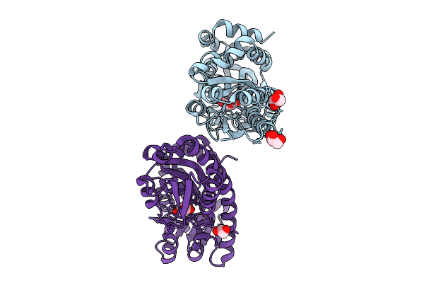 |
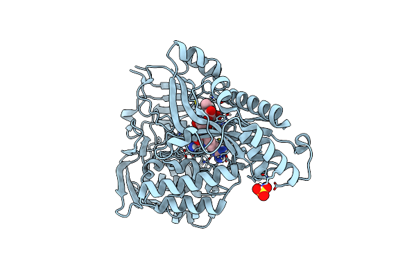
Crystal Structure Of Pa0884, The Sbp Component Of A Pseudomonas Aeruginosa Pao1 Tripartite Atp-Independent Periplasmic (Trap) Transporter, Complexed With Itaconate
Organism: Pseudomonas aeruginosa pao1
Method: X-RAY DIFFRACTION Release Date: 2025-09-10 Classification: TRANSPORT PROTEIN Ligands: ITN, GOL, EDO |
 |
Organism: Leishmania donovani
Method: X-RAY DIFFRACTION Release Date: 2025-07-09 Classification: LIGASE |
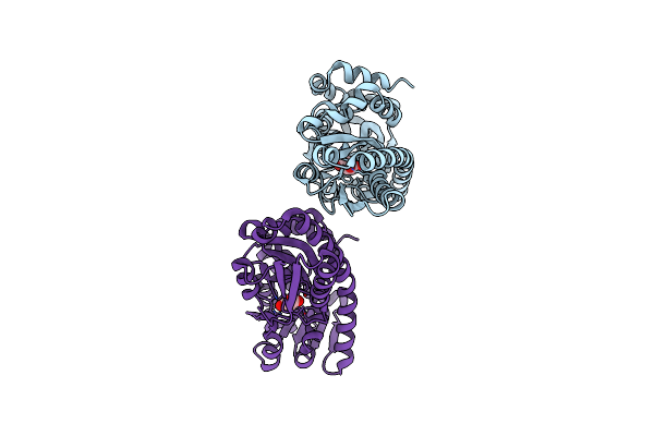 |
Crystal Structure Of Pa0884, The Sbp Component Of A Pseudomonas Aeruginosa Pao1 Tripartite Atp-Independent Periplasmic (Trap) Transporter, Complexed With Succinate
Organism: Pseudomonas aeruginosa pao1
Method: X-RAY DIFFRACTION Release Date: 2025-02-05 Classification: TRANSPORT PROTEIN Ligands: SIN |
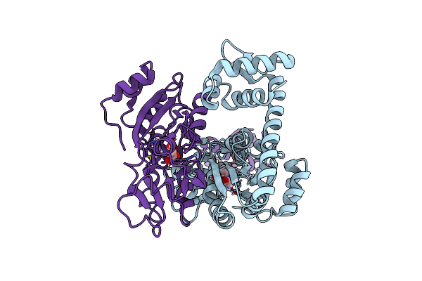 |
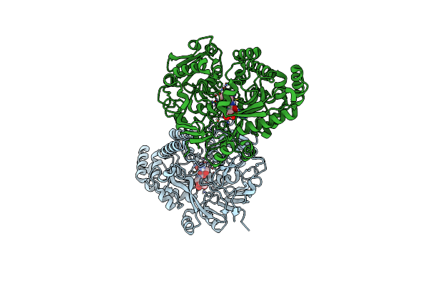
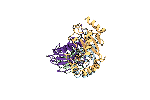
The Crystal Structure Of Full Length Tetramer Cysb From Klebsiella Aerogenes In Complex With N-Acetylserine
Organism: Klebsiella aerogenes
Method: X-RAY DIFFRACTION Resolution:2.30 Å Release Date: 2024-07-24 Classification: TRANSCRIPTION Ligands: SAC |
 |
The Crystal Structure Of Full Length Tetramer Cysb From Klebsiella Aerogenes In Complex With N-Acetylserine
Organism: Klebsiella
Method: X-RAY DIFFRACTION Resolution:2.80 Å Release Date: 2024-07-03 Classification: TRANSCRIPTION Ligands: SAC |
 |
Crystal Structure Of The Second Bromodomain Of Brd5 From Leishmania Donovani
Organism: Leishmania donovani bpk282a1
Method: X-RAY DIFFRACTION Resolution:1.60 Å Release Date: 2023-09-27 Classification: TRANSCRIPTION |
 |
Organism: Haemophilus influenzae
Method: X-RAY DIFFRACTION Resolution:1.80 Å Release Date: 2023-06-14 Classification: RNA BINDING PROTEIN Ligands: NN9, CIT |
 |
Organism: Haemophilus influenzae
Method: X-RAY DIFFRACTION Resolution:1.90 Å Release Date: 2023-06-14 Classification: RNA BINDING PROTEIN Ligands: NMV, CIT |
 |
Organism: Haemophilus influenzae
Method: X-RAY DIFFRACTION Resolution:2.40 Å Release Date: 2023-06-14 Classification: RNA BINDING PROTEIN Ligands: NLL, CIT |
 |
Organism: Haemophilus influenzae
Method: X-RAY DIFFRACTION Resolution:1.80 Å Release Date: 2023-06-14 Classification: RNA BINDING PROTEIN Ligands: CIT, NL1 |
 |
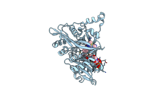
Crystal Structure Of The Peptide Binding Protein, Oppa, From Bacillus Subtilis In Complex With A Phre-Derived Pentapeptide
Organism: Bacillus subtilis subsp. subtilis str. 168
Method: X-RAY DIFFRACTION Resolution:1.90 Å Release Date: 2023-02-22 Classification: TRANSPORT PROTEIN Ligands: SO4 |
 |
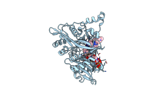
Crystal Structure Of The Peptide Binding Protein, Oppa, From Bacillus Subtilis In Complex With An Endogenous Tetrapeptide
Organism: Bacillus subtilis subsp. subtilis str. 168, Escherichia coli
Method: X-RAY DIFFRACTION Resolution:1.50 Å Release Date: 2023-02-22 Classification: TRANSPORT PROTEIN |
 |
Crystal Structure Of The Peptide Binding Protein Dppe From Bacillus Subtilis In Complex With Murein Tripeptide
Organism: Bacillus subtilis
Method: X-RAY DIFFRACTION Resolution:1.51 Å Release Date: 2023-02-22 Classification: TRANSPORT PROTEIN Ligands: MHI, MG, EDO |
 |
Crystal Structure Of The Peptide Binding Protein Dppe From Bacillus Subtilis In The Unliganded State
Organism: Bacillus subtilis subsp. subtilis str. 168
Method: X-RAY DIFFRACTION Resolution:1.40 Å Release Date: 2023-02-22 Classification: TRANSPORT PROTEIN |
 |
Organism: Escherichia coli (strain k12), Escherichia coli k-12
Method: X-RAY DIFFRACTION Resolution:1.55 Å Release Date: 2021-11-17 Classification: PEPTIDE BINDING PROTEIN Ligands: GOL |
 |
Crystal Structure Of The Chitinase Domain Of The Spore Coat Protein Cote From Clostridium Difficile
Organism: Peptoclostridium difficile (strain 630), Escherichia coli k-12
Method: X-RAY DIFFRACTION Resolution:1.30 Å Release Date: 2020-07-22 Classification: STRUCTURAL PROTEIN Ligands: 1PE, PEG |
 |
The Structure Of An E2 Ubiquitin-Conjugating Complex (Ubc2-Uev1) Essential For Leishmania Amastigote Differentiation
Organism: Leishmania mexicana (strain mhom/gt/2001/u1103)
Method: X-RAY DIFFRACTION Resolution:1.70 Å Release Date: 2020-07-22 Classification: SIGNALING PROTEIN |
 |
Crystal Structure Of The Chitinase Domain Of The Spore Coat Protein Cote From Clostridium Difficile
Organism: Clostridioides difficile (strain 630)
Method: X-RAY DIFFRACTION Resolution:2.10 Å Release Date: 2020-06-24 Classification: STRUCTURAL PROTEIN Ligands: PEG |
 |
Leishmania Major N-Myristoyltransferase In Complex With Thienopyrimidine Inhibitor Imp-0000065
Organism: Leishmania major
Method: X-RAY DIFFRACTION Resolution:1.46 Å Release Date: 2020-05-06 Classification: TRANSFERASE Ligands: MYA, HWT, MG |
 |
Leishmania Major N-Myristoyltransferase In Complex With Quinazoline Inhibitor Imp-0000811
Organism: Leishmania major
Method: X-RAY DIFFRACTION Resolution:1.60 Å Release Date: 2020-05-06 Classification: TRANSFERASE Ligands: MYA, MG, HWZ |