
Search Count: 26
 |
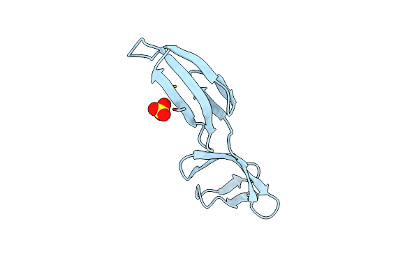
Organism: Homo sapiens
Method: X-RAY DIFFRACTION Resolution:2.20 Å Release Date: 2020-07-29 Classification: STRUCTURAL PROTEIN |
 |
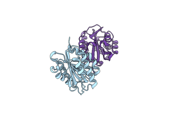
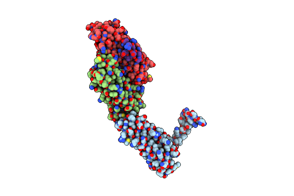
Crystal Structure Of A Disulfide Trapped Single Chain Trimer Composed Of The Mhc I Heavy Chain H-2Kb Y84C K66A Mutant, Beta-2Microglobulin, And Ovalbumin-Derived Peptide
Organism: Mus musculus
Method: X-RAY DIFFRACTION Resolution:2.05 Å Release Date: 2018-04-18 Classification: IMMUNE SYSTEM |
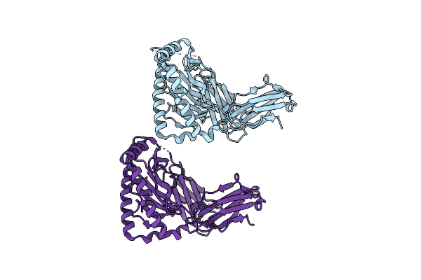 |
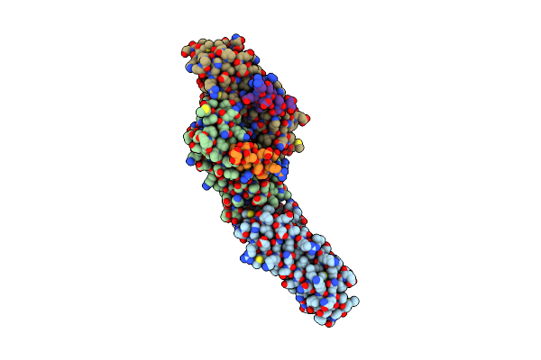
Crystal Structure Of A Single Chain Trimer Composed Of The Mhc 1 Heavy Chain H2-Kb Wt, Beta-2Microglobulin, And Ovalbumin Derived Peptide
Organism: Mus musculus
Method: X-RAY DIFFRACTION Resolution:2.27 Å Release Date: 2018-04-11 Classification: IMMUNE SYSTEM |
 |
Crystal Structure Of A Single Chain Trimer Composed Of The Mhc I Heavy Chain H-2Kb W167A, Beta-2Microglobulin, And And Ovalbumin-Derived Peptide.
Organism: Mus musculus
Method: X-RAY DIFFRACTION Resolution:1.90 Å Release Date: 2018-04-11 Classification: IMMUNE SYSTEM |
 |
Crystal Structure Of A Disulfide Trapped Single Chain Trimer Composed Of The Mhc I Heavy Chain H-2Kb Y84C E63A Mutant, Beta-2Microglobulin, And Ovalbumin-Derived Peptide
Organism: Mus musculus
Method: X-RAY DIFFRACTION Resolution:2.40 Å Release Date: 2018-04-11 Classification: IMMUNE SYSTEM |
 |
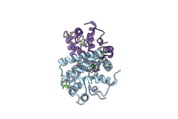
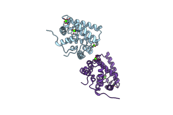
Organism: Streptomyces coelicolor
Method: SOLUTION NMR Release Date: 2016-08-03 Classification: TRANSCRIPTION Ligands: ZN |
 |
Organism: Streptomyces coelicolor
Method: SOLUTION NMR Release Date: 2016-08-03 Classification: TRANSCRIPTION |
 |
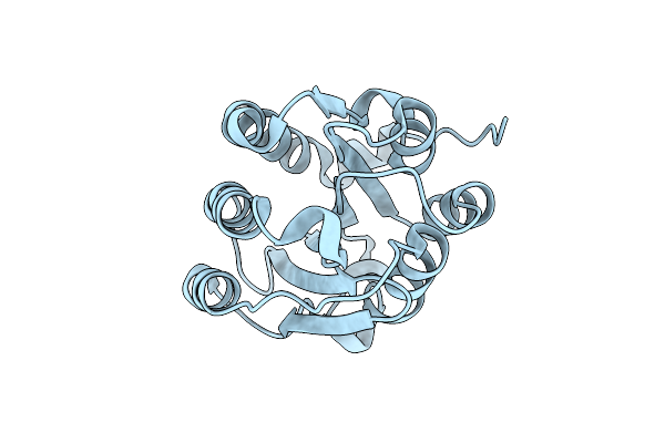
X-Ray Structure Of A Mutant (T61D) Of Calexcitin - A Neuronal Calcium-Signalling Protein
Organism: Doryteuthis pealeii
Method: X-RAY DIFFRACTION Resolution:2.00 Å Release Date: 2014-10-29 Classification: SIGNALING PROTEIN Ligands: CA |
 |
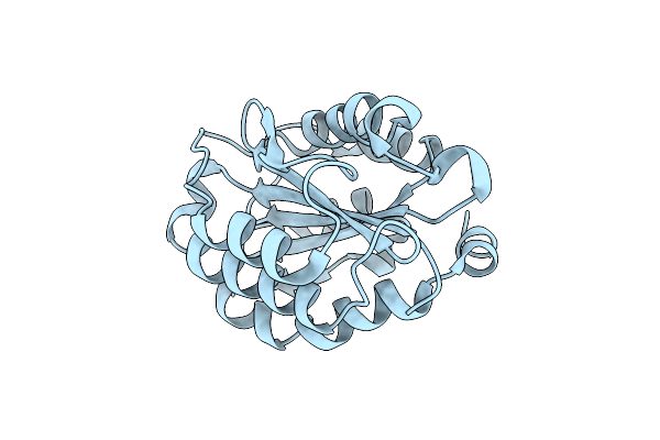
X-Ray Structure Of A Mutant (T188D) Of Calexcitin - A Neuronal Calcium-Signalling Protein
Organism: Doryteuthis pealeii
Method: X-RAY DIFFRACTION Resolution:2.00 Å Release Date: 2014-10-29 Classification: SIGNALING PROTEIN Ligands: CA |
 |
X-Ray Structure Of A Double Mutant Of Calexcitin - A Neuronal Calcium-Signalling Protein
Organism: Doryteuthis pealeii
Method: X-RAY DIFFRACTION Resolution:2.90 Å Release Date: 2014-10-29 Classification: SIGNALING PROTEIN Ligands: CA |
 |
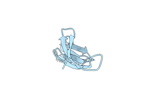
Organism: Mus musculus
Method: X-RAY DIFFRACTION Resolution:1.20 Å Release Date: 2013-12-18 Classification: CELL ADHESION |
 |
Organism: Mus musculus
Method: X-RAY DIFFRACTION Resolution:1.20 Å Release Date: 2013-12-18 Classification: CELL ADHESION |
 |
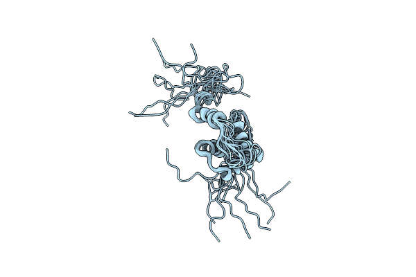
Organism: Homo sapiens
Method: SOLUTION NMR Release Date: 2007-04-03 Classification: SIGNALING PROTEIN |
 |
Organism: Homo sapiens
Method: X-RAY DIFFRACTION Resolution:1.55 Å Release Date: 2007-02-27 Classification: SIGNALING PROTEIN Ligands: SO4 |
 |
Organism: Homo sapiens
Method: X-RAY DIFFRACTION Resolution:1.20 Å Release Date: 2007-02-27 Classification: SIGNALING PROTEIN |
 |
Organism: Loligo pealeii
Method: X-RAY DIFFRACTION Resolution:1.80 Å Release Date: 2006-01-26 Classification: SIGNALING PROTEIN Ligands: CA |
 |
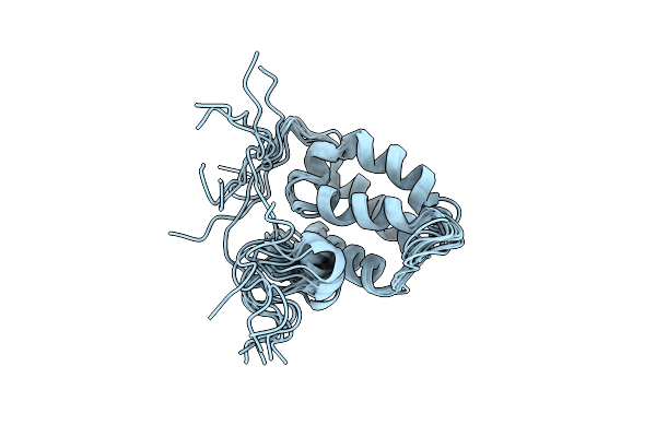
Organism: Homo sapiens
Method: SOLUTION NMR Release Date: 2004-10-12 Classification: SIGNALING PROTEIN |
 |
Paxillin Ld4 Motif Bound To The Focal Adhesion Targeting (Fat) Domain Of The Focal Adhesion Kinase
Organism: Homo sapiens
Method: X-RAY DIFFRACTION Resolution:2.35 Å Release Date: 2003-10-21 Classification: TRANSFERASE |
 |
Paxillin Ld4 Motif Bound To The Focal Adhesion Targeting (Fat) Domain Of The Focal Adhesion Kinase
Organism: Homo sapiens
Method: X-RAY DIFFRACTION Resolution:2.60 Å Release Date: 2003-10-21 Classification: TRANSFERASE |
 |
Paxillin Ld2 Motif Bound To The Focal Adhesion Targeting (Fat) Domain Of The Focal Adhesion Kinase
Organism: Homo sapiens
Method: X-RAY DIFFRACTION Resolution:2.85 Å Release Date: 2003-10-21 Classification: TRANSFERASE |