
Search Count: 26
 |
Organism: Escherichia coli
Method: ELECTRON MICROSCOPY Release Date: 2023-11-29 Classification: DNA BINDING PROTEIN |
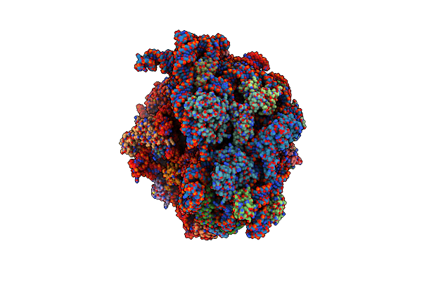 |
Organism: Escherichia coli
Method: ELECTRON MICROSCOPY Release Date: 2023-11-29 Classification: RIBOSOME Ligands: ZN |
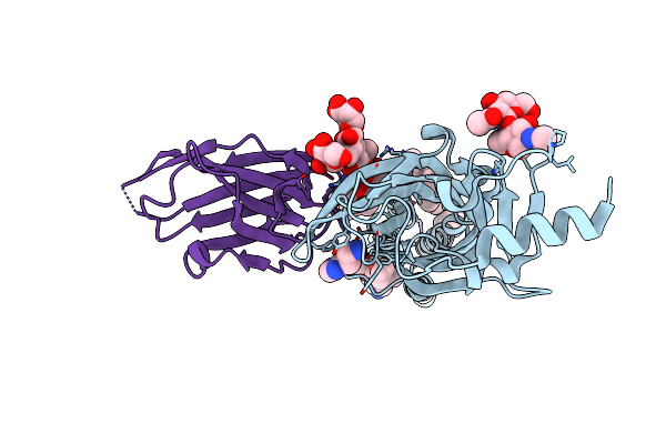 |
Organism: Homo sapiens, Lama glama
Method: ELECTRON MICROSCOPY Release Date: 2023-11-29 Classification: MEMBRANE PROTEIN Ligands: R16, D10, HSM, CL, V8D |
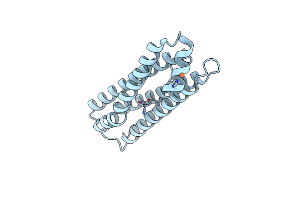 |
Organism: Mus musculus
Method: ELECTRON MICROSCOPY Release Date: 2023-11-29 Classification: METAL BINDING PROTEIN Ligands: FE, ZN |
 |
Organism: Homo sapiens
Method: ELECTRON MICROSCOPY Release Date: 2023-11-29 Classification: OXIDOREDUCTASE Ligands: NDP, HEM |
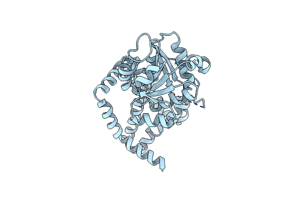 |
Organism: Thermus thermophilus
Method: ELECTRON MICROSCOPY Release Date: 2023-11-29 Classification: OXIDOREDUCTASE |
 |
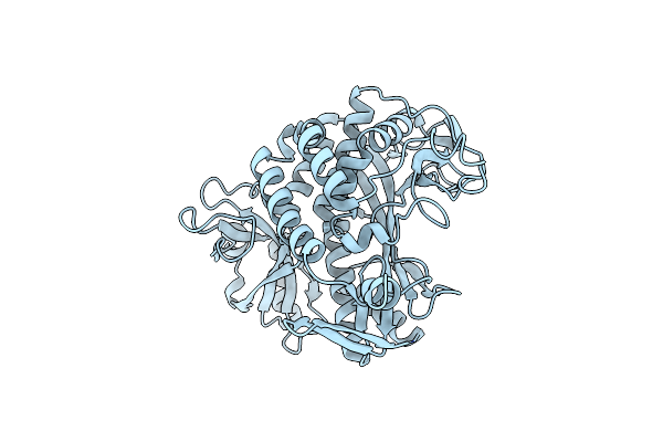
Organism: Cryptosporidium parvum
Method: ELECTRON MICROSCOPY Release Date: 2023-11-29 Classification: OXIDOREDUCTASE Ligands: NAD |
 |
Organism: Escherichia coli
Method: ELECTRON MICROSCOPY Release Date: 2023-11-29 Classification: BIOSYNTHETIC PROTEIN |
 |
Organism: Homo sapiens
Method: ELECTRON MICROSCOPY Release Date: 2023-11-29 Classification: OXIDOREDUCTASE Ligands: CL |
 |
Organism: Escherichia coli
Method: ELECTRON MICROSCOPY Release Date: 2023-11-29 Classification: HYDROLASE |
 |
Organism: Aquifex aeolicus
Method: ELECTRON MICROSCOPY Release Date: 2023-11-29 Classification: TRANSFERASE |
 |
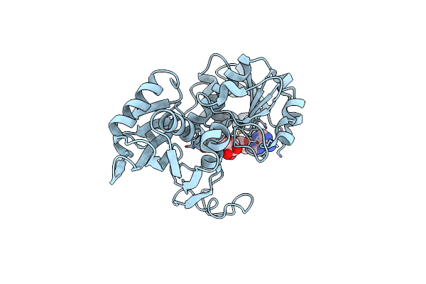
The Transcriptional Regulator Prfa From Listeria Monocytogenes In Complex With Inhibitor Of Wnt Production (Iwp)-2
Organism: Listeria monocytogenes
Method: X-RAY DIFFRACTION Resolution:2.00 Å Release Date: 2021-10-27 Classification: TRANSCRIPTION Ligands: 9XK, DMS, NA |
 |
Organism: Homo sapiens
Method: X-RAY DIFFRACTION Resolution:2.60 Å Release Date: 2021-03-24 Classification: IMMUNE SYSTEM Ligands: GOL |
 |
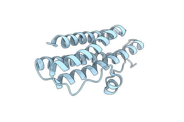
Structure-Function Analyses Of Dual-Bon Domain Protein Dolp Identifies Phospholipid Binding As A New Mechanism For Protein Localisation To The Cell Division Site
Organism: Escherichia coli (strain k12)
Method: SOLUTION NMR Release Date: 2020-12-30 Classification: PROTEIN BINDING |
 |
Organism: Salmonella enterica subsp. enterica serovar typhimurium
Method: X-RAY DIFFRACTION Resolution:1.58 Å Release Date: 2010-03-09 Classification: OXIDOREDUCTASE |
 |
Organism: Salmonella enterica subsp. enterica serovar typhimurium
Method: X-RAY DIFFRACTION Resolution:1.57 Å Release Date: 2010-03-09 Classification: OXIDOREDUCTASE |
 |
Organism: Salmonella enterica subsp. enterica serovar typhimurium
Method: X-RAY DIFFRACTION Resolution:2.15 Å Release Date: 2010-03-09 Classification: OXIDOREDUCTASE Ligands: PE8, P4C, P6G |
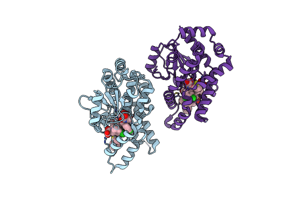 |
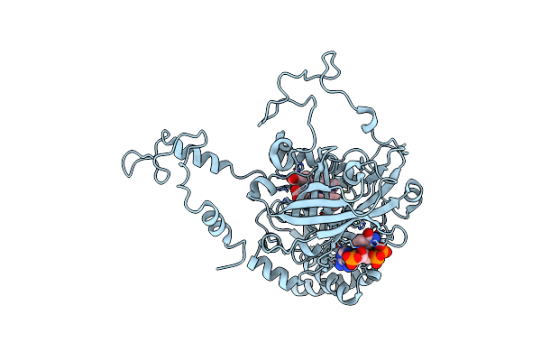
Crystal Structure Of Mycobacterium Tuberculosis Protein Tyrosine Phosphatase Ptpb In Complex With The Specific Inhibitor Omts
Organism: Mycobacterium tuberculosis
Method: X-RAY DIFFRACTION Resolution:2.00 Å Release Date: 2007-05-01 Classification: STRUCTURAL GENOMICS, UNKNOWN FUNCTION Ligands: 7XY |
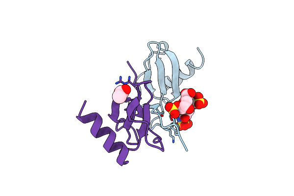 |
Organism: Homo sapiens
Method: X-RAY DIFFRACTION Resolution:2.00 Å Release Date: 2004-11-09 Classification: ATTRACTANT Ligands: ACY |
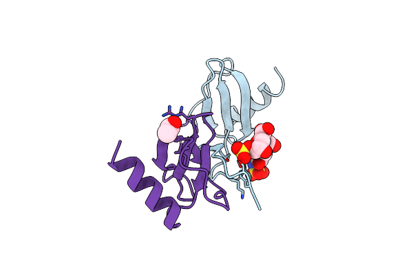 |
Organism: Homo sapiens
Method: X-RAY DIFFRACTION Resolution:2.00 Å Release Date: 2004-11-09 Classification: ATTRACTANT Ligands: ACY |