
Search Count: 3
 |
Organism: Murine coronavirus (strain a59)
Method: X-RAY DIFFRACTION Resolution:3.05 Å Release Date: 2016-02-10 Classification: VIRAL PROTEIN |
 |
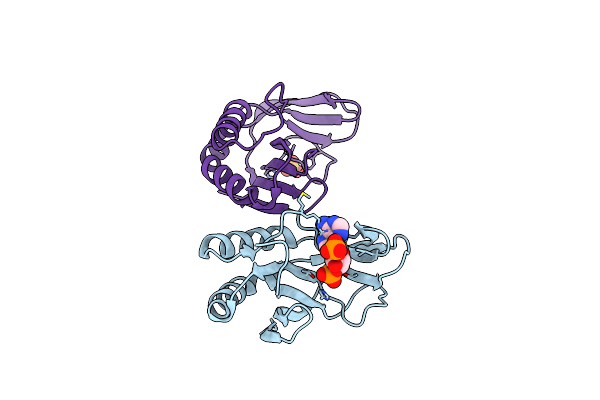
The 1.35A Structure Of A Viral Rnase L Antagonist Reveals Basis For The 2'-5'-Oligoadenylate Binding And Enzyme Activity.
Organism: Rotavirus a
Method: X-RAY DIFFRACTION Resolution:1.35 Å Release Date: 2015-04-29 Classification: VIRAL PROTEIN Ligands: MLI, EDO |
 |
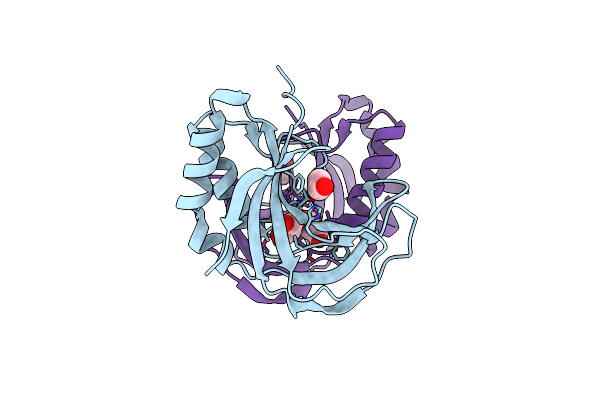
The 1.35 Structure Of A Viral Rnase L Antagonist Reveals Basis For The 2'-5'-Oligoadenylate Binding And Enzyme Activity.
Organism: Rotavirus a
Method: X-RAY DIFFRACTION Resolution:3.10 Å Release Date: 2015-04-29 Classification: VIRAL PROTEIN Ligands: A2P, SO4 |