
Search Count: 6
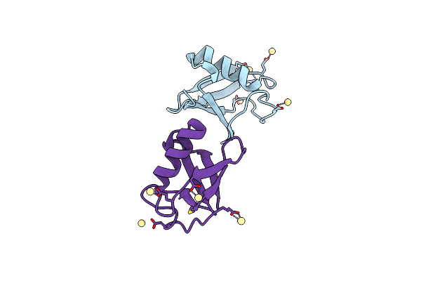 |
Organism: Homo sapiens
Method: X-RAY DIFFRACTION Resolution:2.60 Å Release Date: 2022-05-18 Classification: PROTEIN BINDING Ligands: CD |
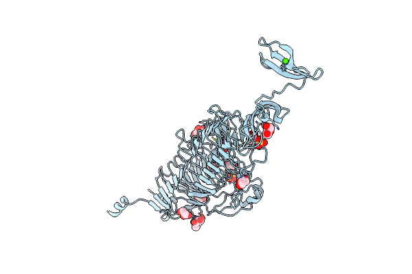 |
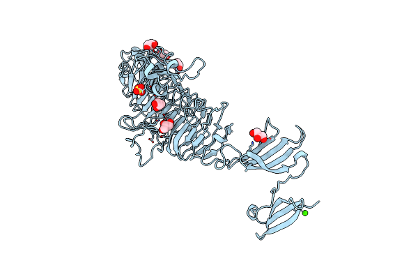
Organism: Enterobacteria phage p22
Method: X-RAY DIFFRACTION Resolution:1.50 Å Release Date: 2008-12-16 Classification: HYDROLASE Ligands: GOL, SO4, CA |
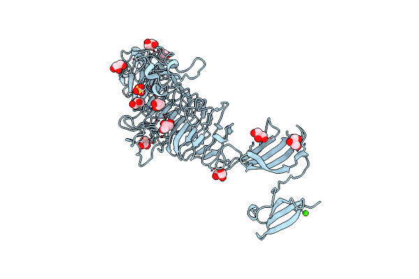 |
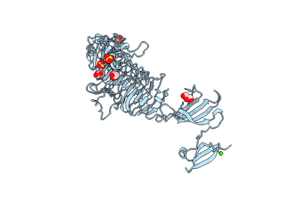
Low Temperature Structure Of P22 Tailspike Protein Fragment (109-666), Mutant V125A
Organism: Enterobacteria phage p22
Method: X-RAY DIFFRACTION Resolution:1.50 Å Release Date: 2008-12-16 Classification: HYDROLASE Ligands: GOL, SO4, CA |
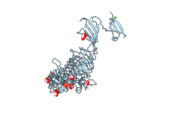 |
Low Temperature Structure Of P22 Tailspike Protein Fragment (109-666), Mutant V125L
Organism: Bacteriophage p22
Method: X-RAY DIFFRACTION Resolution:1.50 Å Release Date: 2008-12-16 Classification: HYDROLASE Ligands: GOL, SO4, CA |
 |
Low Temperature Structure Of P22 Tailspike Protein Fragment (109-666), Mutant V349L
Organism: Enterobacteria phage p22
Method: X-RAY DIFFRACTION Resolution:1.55 Å Release Date: 2008-12-16 Classification: HYDROLASE Ligands: GOL, SO4, CA |
 |
Low Temperature Structure Of P22 Tailspike Protein Fragment (109-666), Mutant V450A
Organism: Enterobacteria phage p22
Method: X-RAY DIFFRACTION Resolution:1.55 Å Release Date: 2008-12-16 Classification: HYDROLASE Ligands: GOL, SO4, CA |