
Search Count: 44
 |
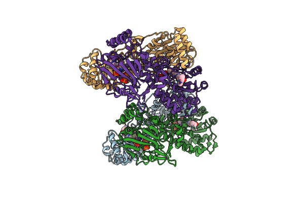
Cryo-Em Structure Of The Human Sk2-4 Chimera/Calmodulin Channel Complex In The Ca2+ Bound State
Organism: Homo sapiens
Method: ELECTRON MICROSCOPY Release Date: 2025-07-09 Classification: TRANSPORT PROTEIN Ligands: K, CA |
 |
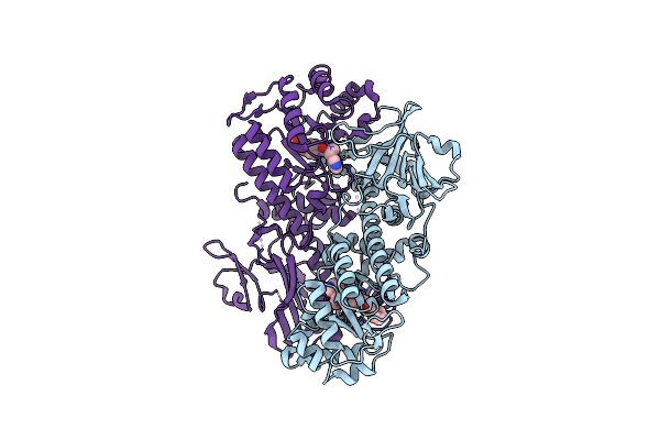
Cryo-Em Structure Of The Human Sk2-4 Chimera/Calmodulin Channel Complex In The Ca2+ Free State
Organism: Homo sapiens
Method: ELECTRON MICROSCOPY Release Date: 2025-07-09 Classification: TRANSPORT PROTEIN Ligands: K, CA |
 |
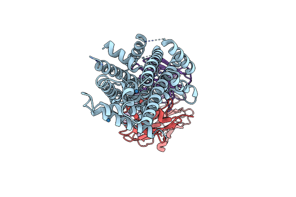
Cryo-Em Structure Of The Human Sk2-4 Chimera/Calmodulin Channel Complex Bound To The Bee Toxin Apamin
Organism: Homo sapiens, Apis mellifera
Method: ELECTRON MICROSCOPY Release Date: 2025-07-09 Classification: TRANSPORT PROTEIN/TOXIN Ligands: K, CA |
 |
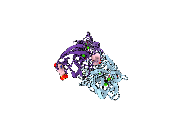
Cryo-Em Structure Of The Human Sk2-4 Chimera/Calmodulin Channel Complex Bound To A Small Molecule Inhibitor
Organism: Homo sapiens
Method: ELECTRON MICROSCOPY Release Date: 2025-07-09 Classification: TRANSPORT PROTEIN/INHIBITOR Ligands: A1B8D, K, CA |
 |
Cryo-Em Structure Of The Human Sk2-4 Chimera/Calmodulin Channel Complex Bound To A Small Molecule Activator
Organism: Homo sapiens
Method: ELECTRON MICROSCOPY Release Date: 2025-07-09 Classification: TRANSPORT PROTEIN Ligands: A1B8G, K, CA |
 |
Structure Of The Voltage-Gated Sodium Channel Navpas From American Cockroach Periplaneta Americana In Complex With Scorpion Alpha-Toxin Lqhait
Organism: Periplaneta americana, Leiurus quinquestriatus hebraeus
Method: ELECTRON MICROSCOPY Release Date: 2024-09-04 Classification: MEMBRANE PROTEIN Ligands: NAG |
 |
Human Plasmakallikrein Protease Domain In Complex With Active Site Directed Inhibitor
Organism: Homo sapiens
Method: X-RAY DIFFRACTION Resolution:1.42 Å Release Date: 2020-07-08 Classification: HYDROLASE Ligands: GSH, DMS, PO4, GOL, MU8 |
 |
Coagulation Factor Xi Protease Domain In Complex With Active Site Inhibitor
Organism: Homo sapiens
Method: X-RAY DIFFRACTION Resolution:1.17 Å Release Date: 2020-07-08 Classification: HYDROLASE Ligands: SO4, DMS, NA, J7B |
 |
Coagulation Factor Xi Protease Domain In Complex With Active Site Inhibitor
Organism: Homo sapiens
Method: X-RAY DIFFRACTION Resolution:1.29 Å Release Date: 2020-07-08 Classification: HYDROLASE Ligands: NW2, SO4, DMS |
 |
Coagulation Factor Xi Protease Domain In Complex With Active Site Inhibitor
Organism: Homo sapiens
Method: X-RAY DIFFRACTION Resolution:1.33 Å Release Date: 2020-07-08 Classification: HYDROLASE Ligands: SO4, DMS, NW5 |
 |
Coagulation Factor Xi Protease Domain In Complex With Active Site Inhibitor
Organism: Homo sapiens
Method: X-RAY DIFFRACTION Resolution:2.63 Å Release Date: 2020-07-08 Classification: HYDROLASE Ligands: NWE |
 |
Organism: Homo sapiens
Method: X-RAY DIFFRACTION Resolution:1.26 Å Release Date: 2020-07-01 Classification: HYDROLASE Ligands: QGS |
 |
Organism: Homo sapiens
Method: X-RAY DIFFRACTION Resolution:2.46 Å Release Date: 2019-12-04 Classification: TRANSFERASE Ligands: OE4, PO4 |
 |
Cleavage And Polyadenylation Specificity Factor Subunit 3 (Cpsf3) In Complex With Nvp-Ltm531
Organism: Homo sapiens
Method: X-RAY DIFFRACTION Resolution:2.49 Å Release Date: 2019-11-27 Classification: HYDROLASE Ligands: ZN, JBG, PO4 |
 |
Organism: Homo sapiens
Method: X-RAY DIFFRACTION Resolution:2.53 Å Release Date: 2018-10-03 Classification: TRANSFERASE Ligands: CVP |
 |
Organism: Homo sapiens
Method: X-RAY DIFFRACTION Resolution:2.44 Å Release Date: 2018-10-03 Classification: TRANSFERASE Ligands: CVJ, PO4 |
 |
Organism: Homo sapiens
Method: X-RAY DIFFRACTION Resolution:2.53 Å Release Date: 2018-09-12 Classification: TRANSFERASE Ligands: BWA, GOL, PO4, DMS |
 |
Organism: Homo sapiens
Method: X-RAY DIFFRACTION Resolution:2.53 Å Release Date: 2018-09-12 Classification: TRANSFERASE Ligands: C5V |
 |
Crystal Structure Of Deinococcus Radiodurans Mnth, An Nramp-Family Transition Metal Transporter
Organism: Deinococcus radiodurans, Mus musculus
Method: X-RAY DIFFRACTION Resolution:3.94 Å Release Date: 2016-11-23 Classification: TRANSPORT PROTEIN/IMMUNE SYSTEM Ligands: OS |
 |
Organism: Mus musculus
Method: X-RAY DIFFRACTION Resolution:1.70 Å Release Date: 2015-10-28 Classification: CELL ADHESION Ligands: CA, CL, MPD, NA, EPE |