
Search Count: 10
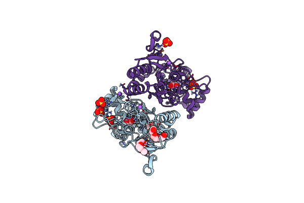 |
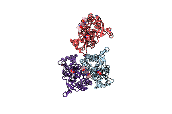
Crystal Structure Of The Glua2 Lbd (L483Y-N754S-L758V) In Complex With Glutamate
Organism: Rattus norvegicus
Method: X-RAY DIFFRACTION Resolution:1.44 Å Release Date: 2017-09-13 Classification: TRANSPORT PROTEIN Ligands: 1PE, PG0, SO4, NA, GLU |
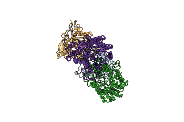 |
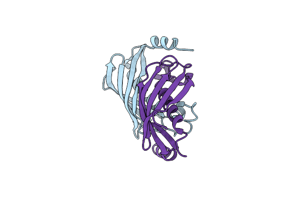
Organism: Treponema denticola atcc 35405
Method: X-RAY DIFFRACTION Resolution:2.00 Å Release Date: 2012-08-29 Classification: OXIDOREDUCTASE |
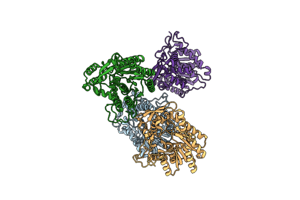 |
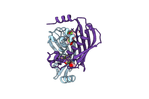
Crystal Structure Of Selenomethionine Containing Trans-2-Enoyl-Coa Reductase From Treponema Denticola
Organism: Treponema denticola
Method: X-RAY DIFFRACTION Resolution:2.05 Å Release Date: 2012-08-29 Classification: OXIDOREDUCTASE |
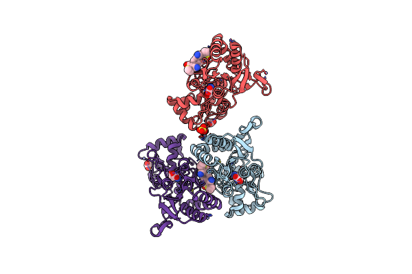 |
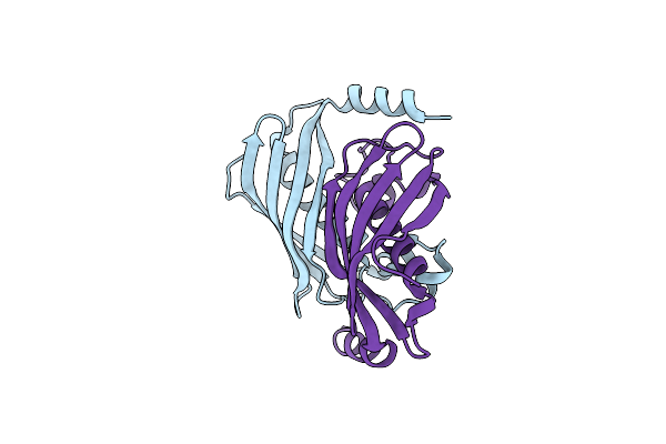
Crystal Structure Of Iglur2 Ligand Binding Domain And Symmetrical Carboxyl Containing Potentiator
Organism: Homo sapiens
Method: X-RAY DIFFRACTION Resolution:1.70 Å Release Date: 2011-05-25 Classification: TRANSPORT PROTEIN Ligands: GLU, RN8, ZN, ACT, SO4 |
 |
Crystal Structure Of Iglur2 Ligand Binding Domain With Symmetric Sulfonamide Containing Potentiator
Organism: Homo sapiens
Method: X-RAY DIFFRACTION Resolution:1.75 Å Release Date: 2011-05-25 Classification: TRANSPORT PROTEIN Ligands: RNN, GLU, ZN |
 |
Organism: Streptomyces cattleya
Method: X-RAY DIFFRACTION Resolution:1.85 Å Release Date: 2010-10-20 Classification: HYDROLASE |
 |
Crystal Structure Of The Fluoroacetyl-Coa-Specific Thioesterase Flk In Complex With Fluoroacetate
Organism: Streptomyces cattleya
Method: X-RAY DIFFRACTION Resolution:2.46 Å Release Date: 2010-10-20 Classification: HYDROLASE Ligands: FAH |
 |
Crystal Structure Of The Fluoroacetyl-Coa-Specific Thioesterase Flk In An Open Conformation
Organism: Streptomyces cattleya
Method: X-RAY DIFFRACTION Resolution:1.95 Å Release Date: 2010-10-20 Classification: HYDROLASE |
 |
Crystal Structure Of The Glur2 Ligand Binding Core (S1S2J) In Complex With Quisqualate And Cx614.
Organism: Rattus norvegicus
Method: X-RAY DIFFRACTION Resolution:1.70 Å Release Date: 2005-10-25 Classification: MEMBRANE PROTEIN Ligands: ZN, QUS, CX6 |
 |
Crystal Structure Of The Glur2 Ligand Binding Core (S1S2J) In Complex With Fluoro-Willardiine And Aniracetam
Organism: Rattus norvegicus
Method: X-RAY DIFFRACTION Resolution:1.65 Å Release Date: 2005-10-25 Classification: MEMBRANE PROTEIN Ligands: FWD, 4MP |