
Search Count: 6
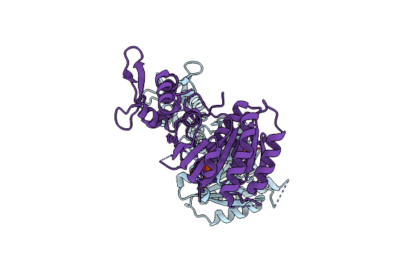 |
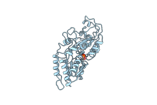
Crystal Structure Of A Dimeric Form Of The Uvsx Recombinase Core Domain From Enterobacteria Phage T4
Organism: Enterobacteria phage t4
Method: X-RAY DIFFRACTION Resolution:2.40 Å Release Date: 2010-08-25 Classification: DNA BINDING PROTEIN Ligands: PO4 |
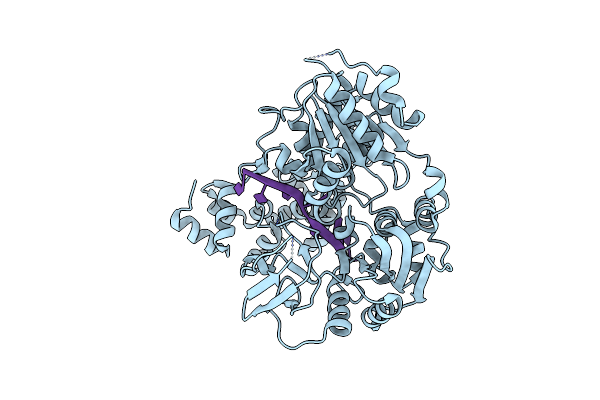 |
Organism: Deinococcus radiodurans r1
Method: X-RAY DIFFRACTION Resolution:2.50 Å Release Date: 2009-06-16 Classification: HYDROLASE/DNA |
 |
Organism: Deinococcus radiodurans r1
Method: X-RAY DIFFRACTION Resolution:2.50 Å Release Date: 2009-06-16 Classification: HYDROLASE/DNA Ligands: MG, ANP |
 |
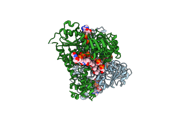
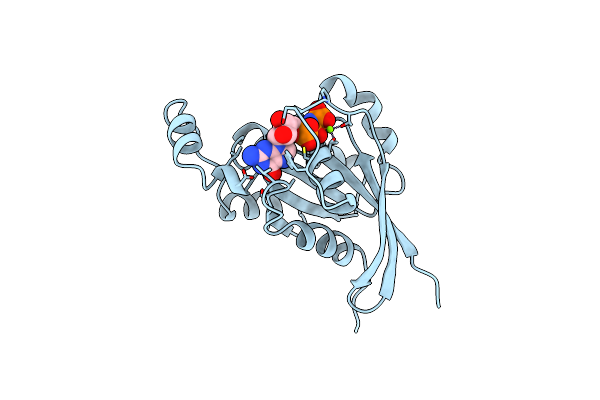
Phosphate-Binding Protein Mutant A197C Labelled With A Coumarin Fluorophore And Bound To Dihydrogenphosphate Ion
Organism: Escherichia coli
Method: X-RAY DIFFRACTION Resolution:1.60 Å Release Date: 1998-10-14 Classification: PHOSPHOTRANSFERASE Ligands: 2HP, MDC |
 |
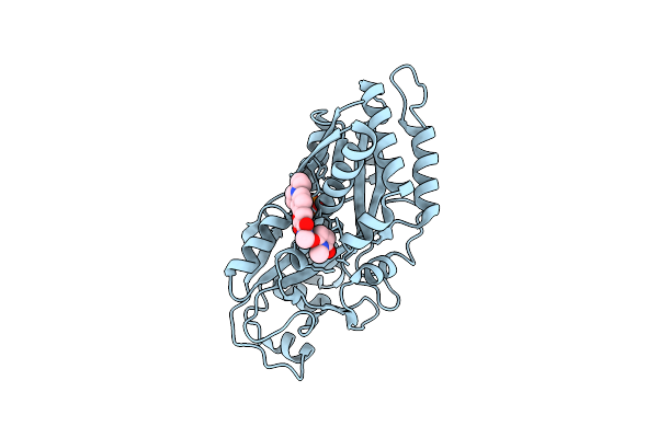
Organism: Escherichia coli
Method: X-RAY DIFFRACTION Resolution:2.40 Å Release Date: 1998-10-14 Classification: PHOSPHOTRANSFERASE Ligands: 2HP |
 |
Organism: Homo sapiens
Method: X-RAY DIFFRACTION Resolution:1.38 Å Release Date: 1998-01-21 Classification: GTP-BINDING Ligands: MG, GNP |