Search Count: 38
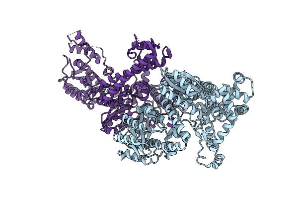 |
Organism: Thermus phage g20c
Method: X-RAY DIFFRACTION Resolution:2.97 Å Release Date: 2022-11-02 Classification: VIRAL PROTEIN |
 |
Organism: Thermus phage tsp4
Method: X-RAY DIFFRACTION Resolution:2.19 Å Release Date: 2022-11-02 Classification: VIRAL PROTEIN Ligands: TMP, MG, TB, CL |
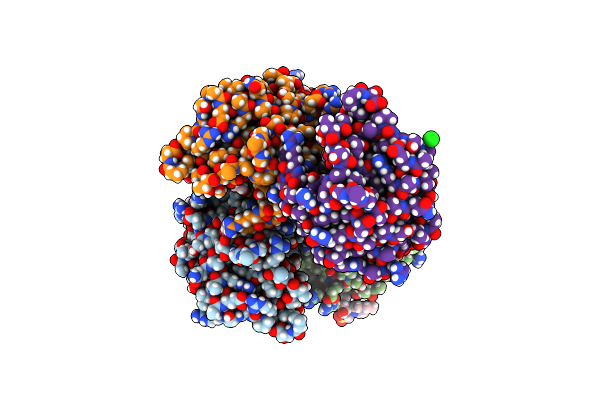 |
Organism: Thermus parvatiensis
Method: X-RAY DIFFRACTION Resolution:1.79 Å Release Date: 2022-06-22 Classification: HYDROLASE Ligands: ZN, GOL, EPE, SO4, NA, CL |
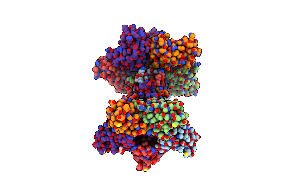 |
Organism: Bacillus subtilis subsp. subtilis str. 168
Method: X-RAY DIFFRACTION Resolution:2.12 Å Release Date: 2019-11-20 Classification: UNKNOWN FUNCTION Ligands: GOL |
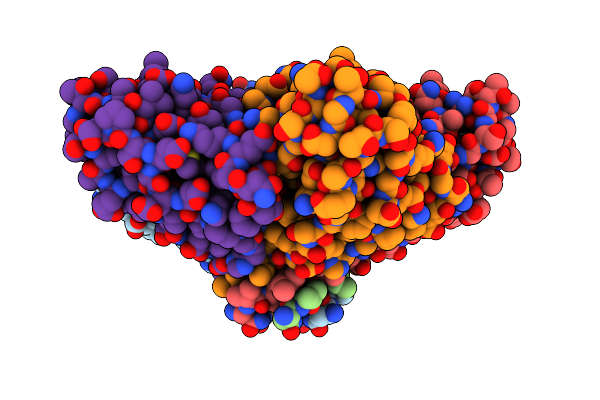 |
Organism: Bacillus subtilis (strain 168)
Method: X-RAY DIFFRACTION Resolution:1.33 Å Release Date: 2019-11-20 Classification: UNKNOWN FUNCTION |
 |
Organism: Bacillus subtilis (strain 168)
Method: X-RAY DIFFRACTION Resolution:1.88 Å Release Date: 2019-11-20 Classification: VIRAL PROTEIN Ligands: GOL, ACT |
 |
Trigonal Crystal Structure Of An Acetylester Hydrolase From Corynebacterium Glutamicum
Organism: Corynebacterium glutamicum
Method: X-RAY DIFFRACTION Resolution:3.20 Å Release Date: 2015-12-16 Classification: HYDROLASE Ligands: GOL |
 |
Organism: Pseudomonas veronii
Method: X-RAY DIFFRACTION Resolution:2.80 Å Release Date: 2015-12-16 Classification: HYDROLASE |
 |
Organism: Pseudomonas veronii
Method: X-RAY DIFFRACTION Resolution:1.82 Å Release Date: 2015-12-16 Classification: HYDROLASE Ligands: EDO, GOL, ACT |
 |
Orthorhombic Crystal Structure Of An Acetylester Hydrolase From Corynebacterium Glutamicum
Organism: Corynebacterium glutamicum (strain atcc 13032 / dsm 20300 / jcm 1318 / lmg 3730 / ncimb 10025)
Method: X-RAY DIFFRACTION Resolution:1.80 Å Release Date: 2015-12-09 Classification: TRANSFERASE Ligands: CL, MG, GOL |
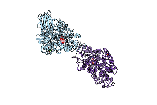 |
Organism: Rhizobium
Method: X-RAY DIFFRACTION Resolution:1.80 Å Release Date: 2013-09-25 Classification: ISOMERASE Ligands: CA, GLC, GOL |
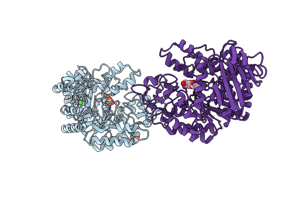 |
Organism: Rhizobium
Method: X-RAY DIFFRACTION Resolution:2.00 Å Release Date: 2013-09-25 Classification: ISOMERASE Ligands: GOL, CA, SO4 |
 |
Mutb Inactive Double Mutant D200A-D415N Soaked With Sucrose And Having As Bound Ligands Sucrose In Molecule A And The Reaction Product Trehalulose In Molecule B
Organism: Rhizobium
Method: X-RAY DIFFRACTION Resolution:2.00 Å Release Date: 2013-09-25 Classification: ISOMERASE Ligands: GOL, CA, TEU |
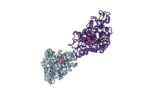 |
Crystal Structure Of The Trehalulose Synthase Mutb In Complex With Trehalulose
Organism: Rhizobium
Method: X-RAY DIFFRACTION Resolution:1.95 Å Release Date: 2013-09-25 Classification: ISOMERASE Ligands: CA, TEU |
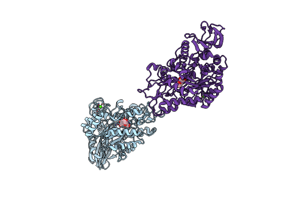 |
Organism: Rhizobium
Method: X-RAY DIFFRACTION Resolution:2.20 Å Release Date: 2013-09-25 Classification: ISOMERASE Ligands: CA, ISL, GLC |
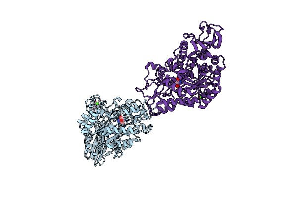 |
Crystal Structure Of The Trehalulose Synthase Mutb, Mutant A258V, In Complex With Tris
Organism: Pseudomonas mesoacidophila
Method: X-RAY DIFFRACTION Resolution:2.15 Å Release Date: 2013-08-21 Classification: ISOMERASE Ligands: CA, TRS |
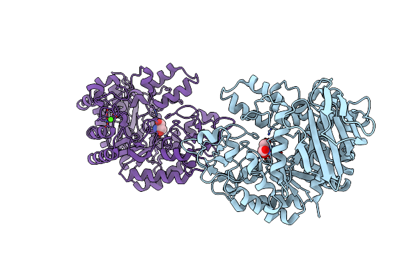 |
Crystal Structure Of The Trehalulose Synthase Mutant, Mutb D415N, In Complex With Tris
Organism: Pseudomonas mesoacidophila
Method: X-RAY DIFFRACTION Resolution:2.20 Å Release Date: 2013-08-21 Classification: ISOMERASE Ligands: CA, TRS |
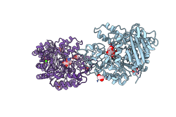 |
Organism: Rhizobium
Method: X-RAY DIFFRACTION Resolution:2.15 Å Release Date: 2013-02-13 Classification: ISOMERASE Ligands: CA, GLC, GOL, TRS |
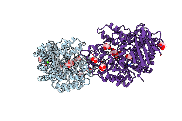 |
Crystal Structure Of The Mutb F164L Mutant From Crystals Soaked With The Substrate Sucrose
Organism: Rhizobium
Method: X-RAY DIFFRACTION Resolution:1.95 Å Release Date: 2013-02-13 Classification: ISOMERASE Ligands: CA, GOL, TRS |
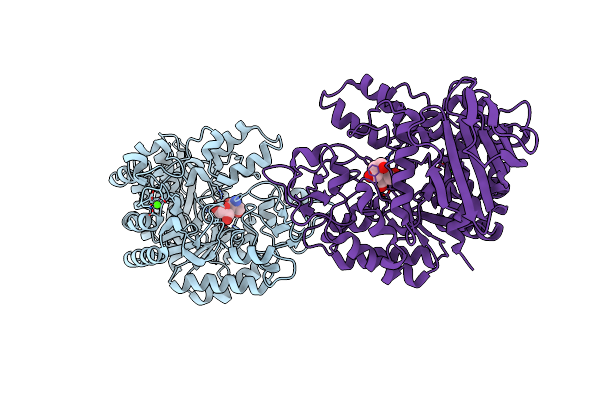 |
Crystal Structure Of The Mutb F164L Mutant From Crystals Soaked With Trehalulose
Organism: Rhizobium
Method: X-RAY DIFFRACTION Resolution:2.15 Å Release Date: 2013-02-13 Classification: ISOMERASE Ligands: CA, TRS, GOL |

