Planned Maintenance: Some services may turn out to be unavailable from 15th January, 2026 to 16th January, 2026. We apologize for the inconvenience!

Planned Maintenance: Some services may turn out to be unavailable from 15th January, 2026 to 16th January, 2026. We apologize for the inconvenience!
 |
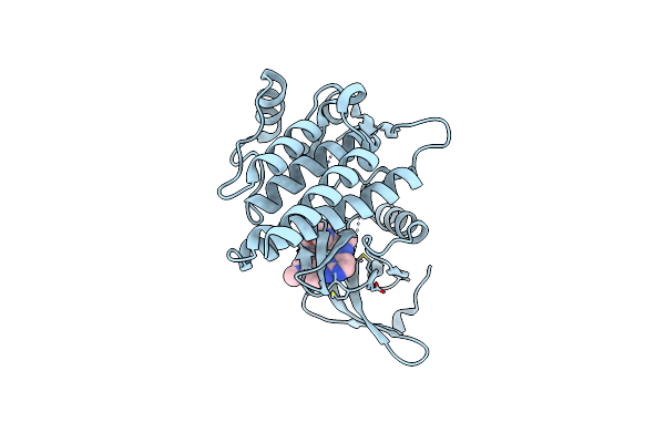
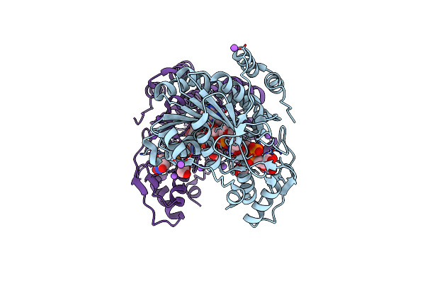
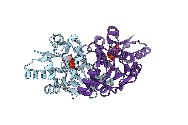
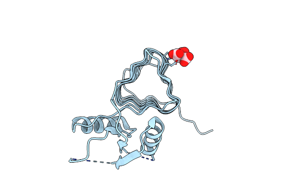
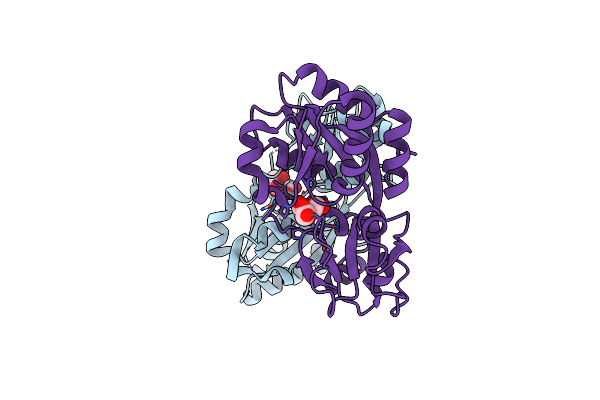
Crystal Structure Of Human Mat2A Bound To S-Adenosylmethionine And Compound 8
Organism: Homo sapiens
Method: X-RAY DIFFRACTION Resolution:1.19 Å Release Date: 2024-03-20 Classification: TRANSFERASE Ligands: U74, SAM |
 |
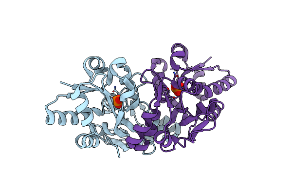
Crystal Structure Of Human Mat2A Bound To S-Adenosylmethionine And Compound 11
Organism: Homo sapiens
Method: X-RAY DIFFRACTION Resolution:1.16 Å Release Date: 2024-03-20 Classification: TRANSFERASE Ligands: U6R, SAM |
 |
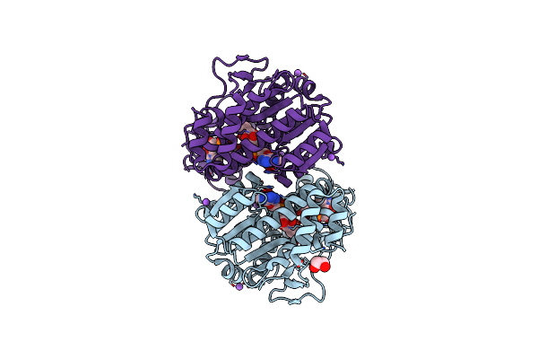
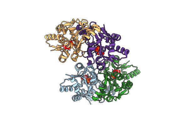
Crystal Structure Of Human Mat2A Bound To S-Adenosylmethionine And Compound 12
Organism: Homo sapiens
Method: X-RAY DIFFRACTION Resolution:1.12 Å Release Date: 2024-03-20 Classification: TRANSFERASE Ligands: U8R, SAM |
 |
Crystal Structure Of Human Mat2A Bound To S-Adenosylmethionine And Compound 15
Organism: Homo sapiens
Method: X-RAY DIFFRACTION Resolution:1.10 Å Release Date: 2024-03-20 Classification: TRANSFERASE Ligands: U8E, SAM |
 |
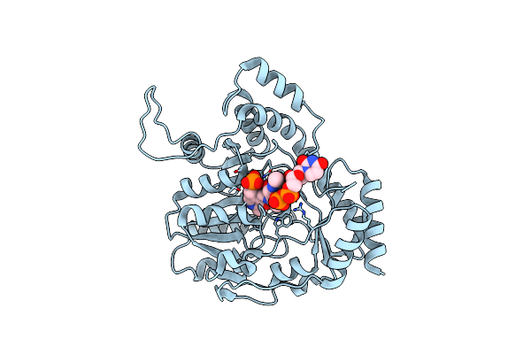
Crystal Structure Of Human Mat2A Bound To S-Adenosylmethionine And Compound 21
Organism: Homo sapiens
Method: X-RAY DIFFRACTION Resolution:1.11 Å Release Date: 2024-03-20 Classification: TRANSFERASE Ligands: U7X, SAM |
 |
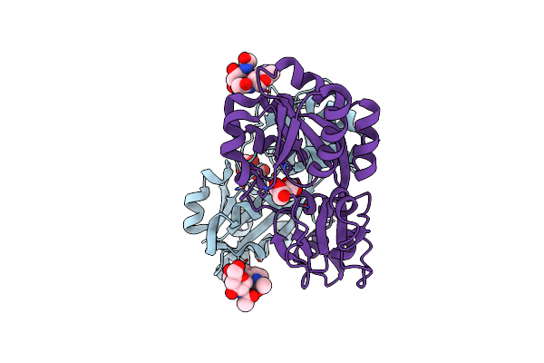
Crystal Structure Of Human Mat2A Bound To S-Adenosylmethionine And Compound 31
Organism: Homo sapiens
Method: X-RAY DIFFRACTION Resolution:1.09 Å Release Date: 2024-03-20 Classification: TRANSFERASE Ligands: U96, SAM |
 |
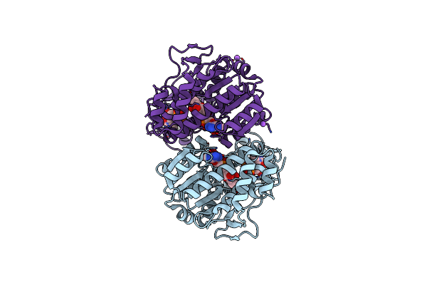
Organism: Homo sapiens
Method: X-RAY DIFFRACTION Resolution:1.51 Å Release Date: 2023-06-14 Classification: TRANSFERASE Ligands: QR7 |
 |
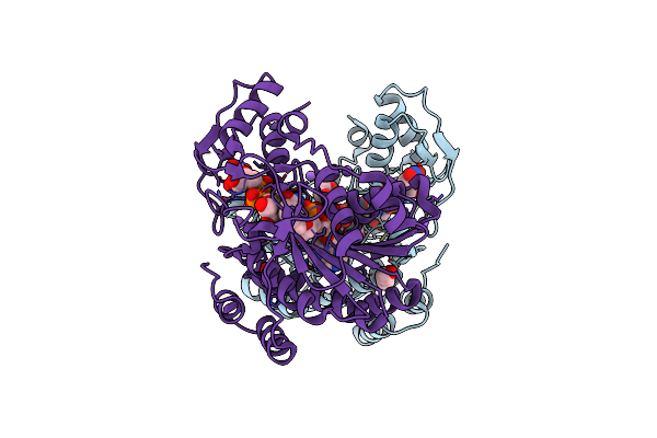
X-Ray Structure Of The Pglf Dehydratase From Campylobacter Jejuni In Complex With Udp And Nad(H)
Organism: Campylobacter jejuni
Method: X-RAY DIFFRACTION Resolution:2.00 Å Release Date: 2017-11-08 Classification: MEMBRANE PROTEIN Ligands: UDP, NAD, EDO, NA |
 |
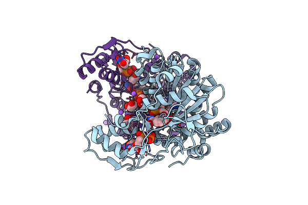
X-Ray Structure Of The Pglf Udp-N-Acetylglucosamine 4,6-Dehydratase From Campylobacterjejuni, D396N/K397A Variant In Complex With Udp-N-Acrtylglucosamine
Organism: Campylobacter jejuni
Method: X-RAY DIFFRACTION Resolution:1.80 Å Release Date: 2017-11-08 Classification: MEMBRANE PROTEIN Ligands: NAD, UD1, EDO, NA |
 |
X-Ray Structure Of The Pglf 4,6-Dehydratase From Campylobacter Jejuni, T595S Variant, In Complex With Udp
Organism: Campylobacter jejuni
Method: X-RAY DIFFRACTION Resolution:1.60 Å Release Date: 2017-11-08 Classification: MEMBRANE PROTEIN Ligands: UDP, NAD, EDO, NA |
 |
X-Ray Structure Of The Pglf 4,6-Dehydratase From Campylobacter Jejuni, Variant T395V, In Complex With Udp
Organism: Campylobacter jejuni
Method: X-RAY DIFFRACTION Resolution:1.60 Å Release Date: 2017-11-08 Classification: MEMBRANE PROTEIN Ligands: UDP, NAD, EDO, NA |
 |
X-Ray Structure Of The Pglf 4,5-Dehydratase From Campylobacter Jejuni, Variant M405Y, In Complex With Udp
Organism: Campylobacter jejuni
Method: X-RAY DIFFRACTION Resolution:1.60 Å Release Date: 2017-11-08 Classification: MEMBRANE PROTEIN Ligands: UDP, NAD, EDO, NA |
 |
Organism: Campylobacter jejuni
Method: X-RAY DIFFRACTION Resolution:2.00 Å Release Date: 2015-07-29 Classification: TRANSFERASE Ligands: 4RA |
 |
Organism: Campylobacter jejuni
Method: X-RAY DIFFRACTION Resolution:2.00 Å Release Date: 2009-03-10 Classification: PROTEIN BINDING Ligands: FLC |
 |
Organism: Campylobacter jejuni
Method: X-RAY DIFFRACTION Resolution:1.70 Å Release Date: 2009-03-10 Classification: PROTEIN BINDING Ligands: PEQ |
 |
Organism: Campylobacter jejuni
Method: X-RAY DIFFRACTION Resolution:2.20 Å Release Date: 2009-03-10 Classification: TRANSPORT PROTEIN Ligands: 3PG |
 |
Organism: Campylobacter jejuni
Method: X-RAY DIFFRACTION Resolution:1.60 Å Release Date: 2009-03-10 Classification: TRANSPORT PROTEIN Ligands: PO4 |
 |
Organism: Campylobacter jejuni
Method: X-RAY DIFFRACTION Resolution:1.80 Å Release Date: 2008-01-29 Classification: TRANSFERASE Ligands: COA, SO4 |
 |
Organism: Campylobacter jejuni
Method: X-RAY DIFFRACTION Resolution:1.75 Å Release Date: 2008-01-22 Classification: TRANSFERASE Ligands: FLC |
 |
Organism: Campylobacter jejuni
Method: X-RAY DIFFRACTION Resolution:1.60 Å Release Date: 2007-05-01 Classification: periplasmic binding protein Ligands: FLC |