
Search Count: 7
 |
Organism: Roystonea regia
Method: X-RAY DIFFRACTION Resolution:1.85 Å Release Date: 2009-11-24 Classification: OXIDOREDUCTASE Ligands: HEM, CA, PEO, SO4, MES, EDO, NAG |
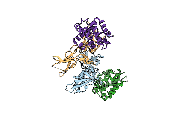 |
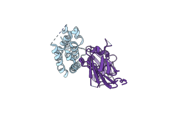
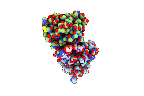
Crystal Structure Of A Soluble Decoy Receptor Il-22Bp Bound To Interleukin-22
Organism: Homo sapiens
Method: X-RAY DIFFRACTION Resolution:2.76 Å Release Date: 2009-04-14 Classification: CYTOKINE/CYTOKINE RECEPTOR |
 |
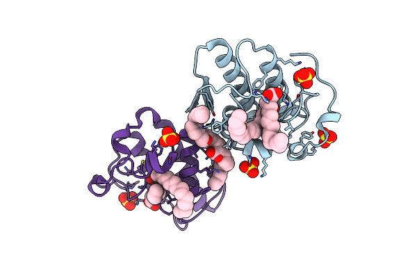
Organism: Homo sapiens
Method: X-RAY DIFFRACTION Resolution:1.90 Å Release Date: 2008-08-19 Classification: CYTOKINE/CYTOKINE RECEPTOR |
 |
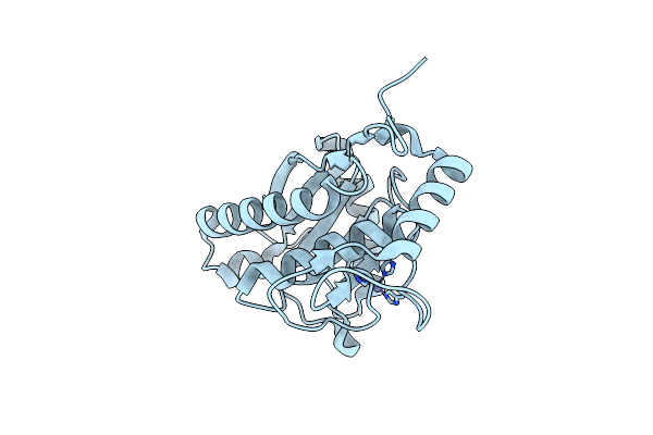
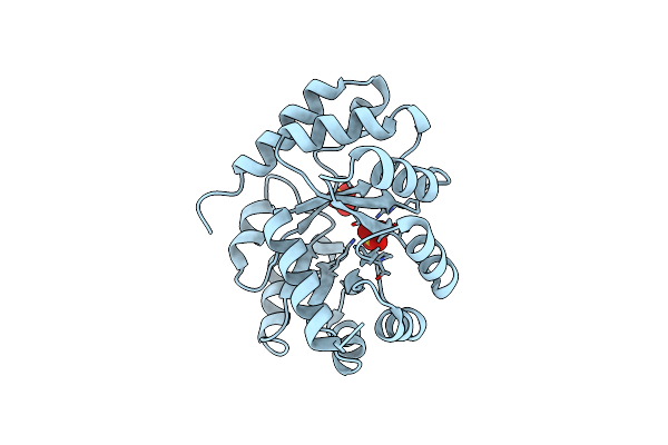
Structural Insights For Fatty Acid Binding In A Lys49 Phospholipase A2: Crystal Structure Of Myotoxin Ii From Bothrops Moojeni Complexed With Stearic Acid
Organism: Bothrops moojeni
Method: X-RAY DIFFRACTION Resolution:1.80 Å Release Date: 2005-03-29 Classification: HYDROLASE Ligands: SO4, STE |
 |
Amino Acid Sequence And Crystal Structure Of Bap1, A Metalloproteinase From Bothrops Asper Snake Venom That Exerts Multiple Tissue-Damaging Activities.
Organism: Bothrops asper
Method: X-RAY DIFFRACTION Resolution:1.93 Å Release Date: 2003-11-04 Classification: TOXIN Ligands: ZN |
 |
Crystal Structure Of 2-Keto-3-Deoxy-6-Phosphogluconate (Kdpg) Aldolase From Pseudomonas Putida.
Organism: Pseudomonas putida
Method: X-RAY DIFFRACTION Resolution:2.20 Å Release Date: 2003-09-16 Classification: LYASE Ligands: SO4 |
 |
Three-Dimensional Structure Of Ribonulcease T1 Complexed With An Isosteric Phosphonate Analogue Of Gpu: Alternate Substrate Binding Modes And Catalysis.
Organism: Aspergillus oryzae
Method: X-RAY DIFFRACTION Resolution:2.00 Å Release Date: 1999-03-25 Classification: HYDROLASE/RNA |