
Search Count: 29
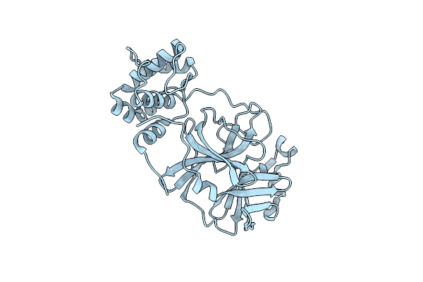 |
Organism: Severe acute respiratory syndrome coronavirus 2
Method: X-RAY DIFFRACTION Resolution:1.60 Å Release Date: 2020-12-23 Classification: VIRAL PROTEIN, HYDROLASE |
 |
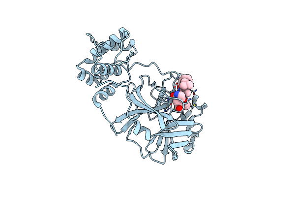
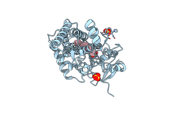
Organism: Severe acute respiratory syndrome coronavirus 2
Method: X-RAY DIFFRACTION Resolution:1.60 Å Release Date: 2020-12-23 Classification: VIRAL PROTEIN, HYDROLASE/INHIBITOR Ligands: GHX |
 |
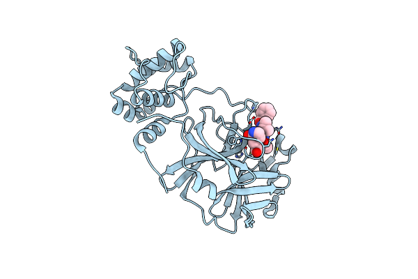
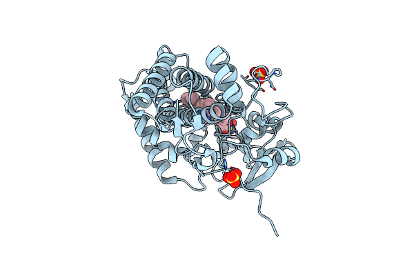
Organism: Severe acute respiratory syndrome coronavirus 2
Method: X-RAY DIFFRACTION Resolution:1.65 Å Release Date: 2020-12-23 Classification: VIRAL PROTEIN, HYDROLASE/INHIBITOR Ligands: VHV |
 |
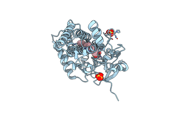
Organism: Severe acute respiratory syndrome coronavirus 2
Method: X-RAY DIFFRACTION Resolution:1.65 Å Release Date: 2020-12-23 Classification: VIRAL PROTEIN, HYDROLASE/INHIBITOR Ligands: VHJ |
 |
Organism: Severe acute respiratory syndrome coronavirus 2
Method: X-RAY DIFFRACTION Resolution:1.40 Å Release Date: 2020-12-23 Classification: VIRAL PROTEIN, HYDROLASE/INHIBITOR Ligands: VHM |
 |
Organism: Severe acute respiratory syndrome coronavirus 2
Method: X-RAY DIFFRACTION Resolution:2.10 Å Release Date: 2020-12-23 Classification: VIRAL PROTEIN, HYDROLASE/INHIBITOR Ligands: VHP |
 |
Organism: Severe acute respiratory syndrome coronavirus 2
Method: X-RAY DIFFRACTION Resolution:1.65 Å Release Date: 2020-12-23 Classification: VIRAL PROTEIN, HYDROLASE/INHIBITOR Ligands: XM2 |
 |
Organism: Severe acute respiratory syndrome coronavirus 2
Method: X-RAY DIFFRACTION Resolution:1.90 Å Release Date: 2020-12-23 Classification: VIRAL PROTEIN, HYDROLASE/INHIBITOR Ligands: NOL |
 |
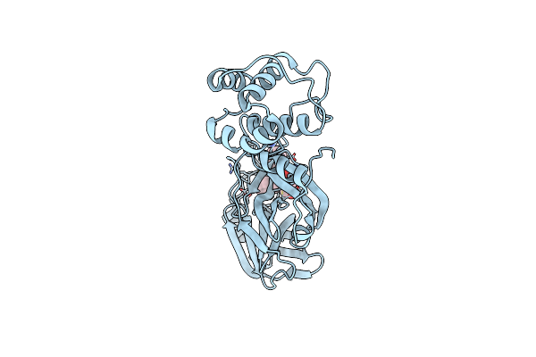
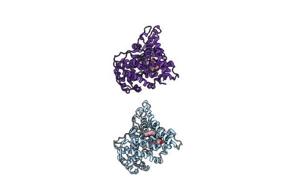
Organism: Bacillus megaterium
Method: X-RAY DIFFRACTION Resolution:2.30 Å Release Date: 2008-12-30 Classification: OXIDOREDUCTASE Ligands: HEM |
 |
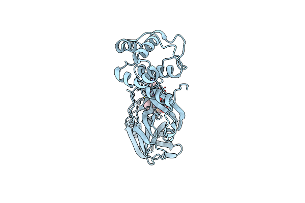
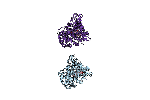
Organism: Bacillus megaterium
Method: X-RAY DIFFRACTION Resolution:2.50 Å Release Date: 2008-12-30 Classification: OXIDOREDUCTASE Ligands: PAM, HEM |
 |
Organism: Bacillus megaterium
Method: X-RAY DIFFRACTION Resolution:2.10 Å Release Date: 2008-12-30 Classification: OXIDOREDUCTASE Ligands: HEM |
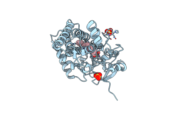 |
Crystal Structure Of The Cytochrome P450 Cyp121 A233G Mutant From Mycobacterium Tuberculosis
Organism: Mycobacterium tuberculosis
Method: X-RAY DIFFRACTION Resolution:1.70 Å Release Date: 2008-09-23 Classification: OXIDOREDUCTASE Ligands: SO4, HEM |
 |
Organism: Mycobacterium tuberculosis
Method: X-RAY DIFFRACTION Resolution:1.90 Å Release Date: 2008-09-23 Classification: OXIDOREDUCTASE Ligands: SO4, HEM |
 |
Crystal Structure Of The Cytochrome P450 Cyp121 P346L Mutant From M. Tuberculosis
Organism: Mycobacterium tuberculosis h37rv
Method: X-RAY DIFFRACTION Resolution:1.45 Å Release Date: 2008-09-23 Classification: OXIDOREDUCTASE Ligands: SO4, HEM |
 |
Crystal Structure Of Cytochrome P450 Cyp121 R386L Mutant From M. Tuberculosis
Organism: Mycobacterium tuberculosis
Method: X-RAY DIFFRACTION Resolution:1.08 Å Release Date: 2008-09-23 Classification: OXIDOREDUCTASE Ligands: SO4, HEM |
 |
Crystal Structure Of Cytochrome P450 Cyp121 S237A Mutant From Mycobacterium Tuberculosis
Organism: Mycobacterium tuberculosis
Method: X-RAY DIFFRACTION Resolution:1.90 Å Release Date: 2008-09-23 Classification: OXIDOREDUCTASE Ligands: SO4, HEM |
 |
Crystal Structure Of The Cytochrome P450 Cyp121 S279A Mutant From M. Tuberculosis
Organism: Mycobacterium tuberculosis
Method: X-RAY DIFFRACTION Resolution:1.75 Å Release Date: 2008-09-23 Classification: OXIDOREDUCTASE Ligands: SO4, HEM |
 |
Organism: Bacillus megaterium
Method: X-RAY DIFFRACTION Resolution:1.20 Å Release Date: 2006-11-07 Classification: OXIDOREDUCTASE Ligands: HEM |
 |
Organism: Bacillus megaterium
Method: X-RAY DIFFRACTION Resolution:1.90 Å Release Date: 2006-11-07 Classification: OXIDOREDUCTASE Ligands: HEM |
 |
Organism: Bacillus megaterium
Method: X-RAY DIFFRACTION Resolution:2.40 Å Release Date: 2006-11-07 Classification: OXIDOREDUCTASE Ligands: HEM |