Planned Maintenance: Some services may turn out to be unavailable from 15th January, 2026 to 16th January, 2026. We apologize for the inconvenience!

Planned Maintenance: Some services may turn out to be unavailable from 15th January, 2026 to 16th January, 2026. We apologize for the inconvenience!
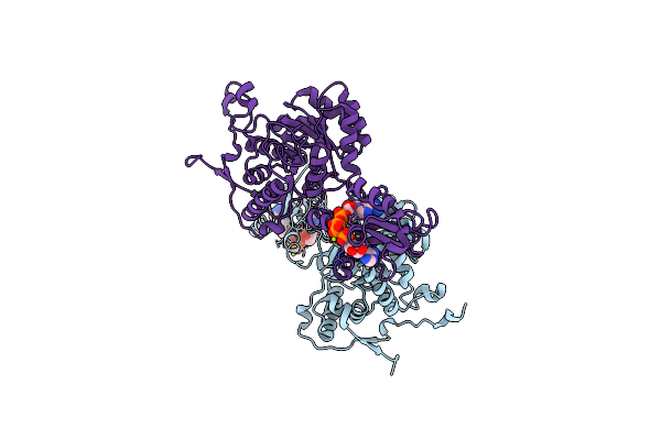 |
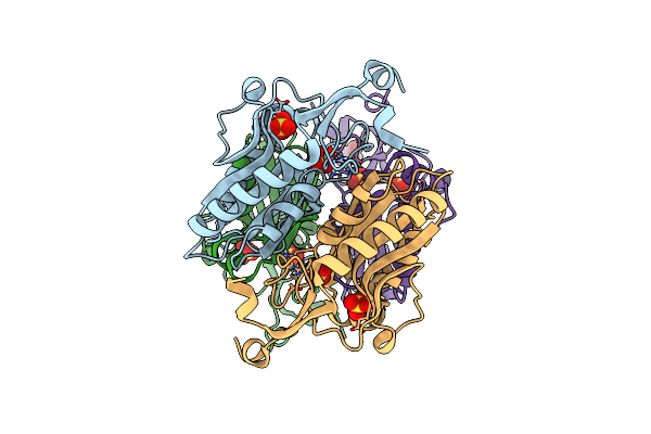
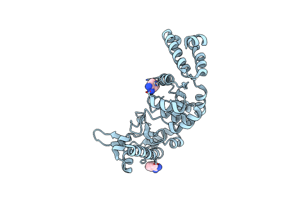
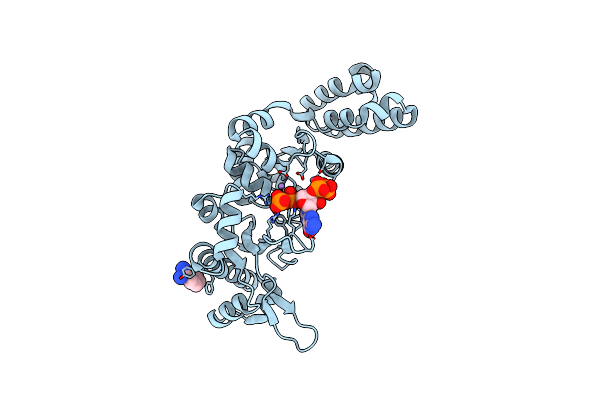
Organism: Bacillus subtilis
Method: X-RAY DIFFRACTION Resolution:2.44 Å Release Date: 2022-09-14 Classification: SIGNALING PROTEIN Ligands: B4P, MG |
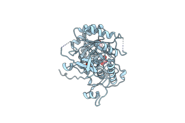 |
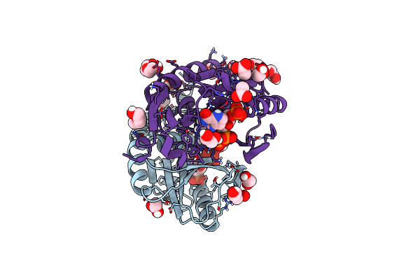
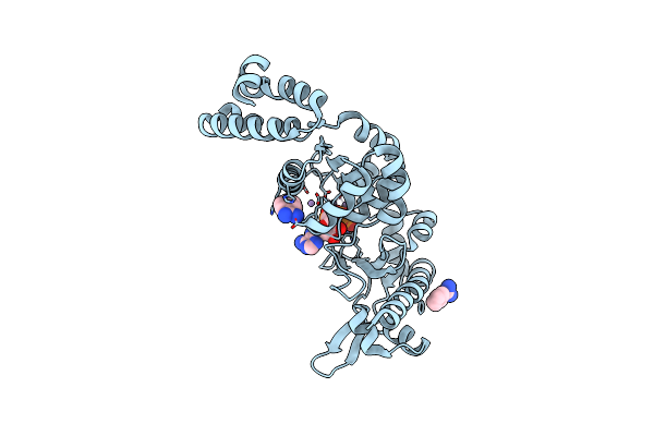
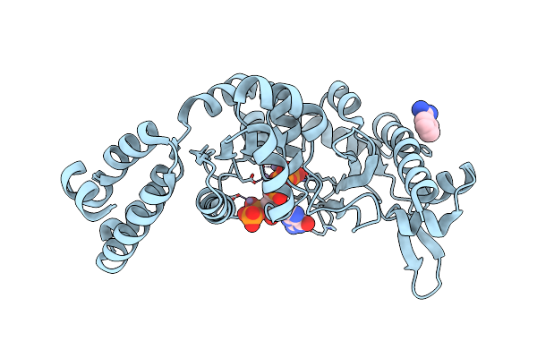
Organism: Bacillus subtilis (strain 168)
Method: X-RAY DIFFRACTION Resolution:1.76 Å Release Date: 2022-09-14 Classification: SIGNALING PROTEIN Ligands: PO4, GOL |
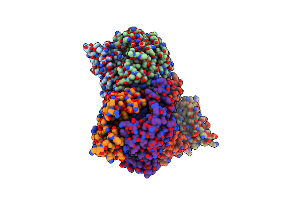 |
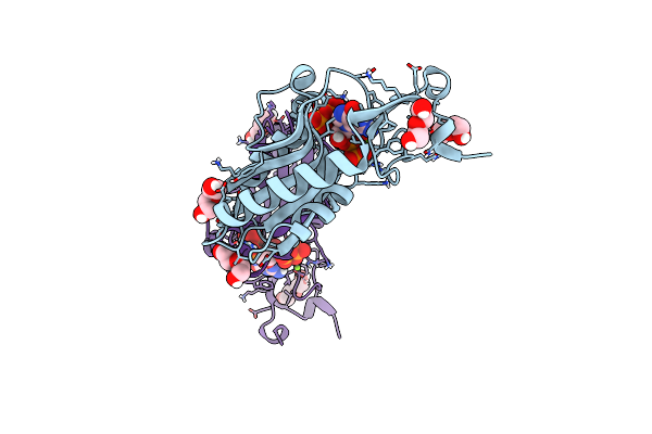
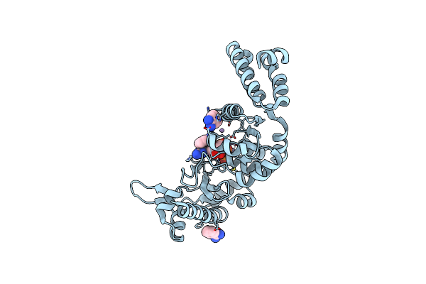
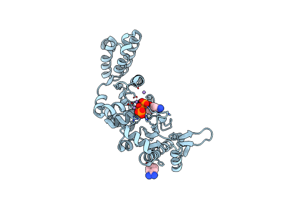
Organism: Bacillus subtilis
Method: X-RAY DIFFRACTION Resolution:2.45 Å Release Date: 2021-12-22 Classification: DNA BINDING PROTEIN Ligands: G4P |
 |
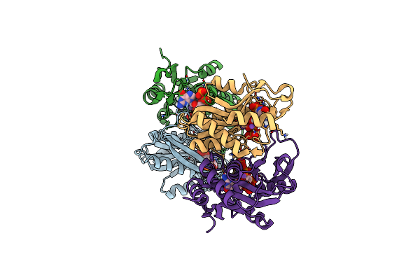
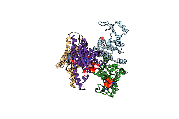
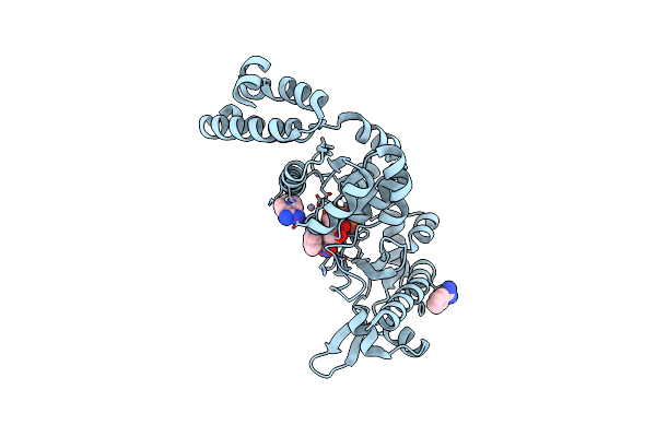
Organism: Listeria monocytogenes
Method: X-RAY DIFFRACTION Resolution:1.62 Å Release Date: 2020-09-09 Classification: CYTOSOLIC PROTEIN Ligands: CL |
 |
Crystal Structure Of Listeria Monocytogenes Cbpb Protein (Lmo1009) In Complex With C-Di-Amp
Organism: Listeria monocytogenes
Method: X-RAY DIFFRACTION Resolution:2.40 Å Release Date: 2020-09-09 Classification: CYTOSOLIC PROTEIN Ligands: 2BA |
 |
Re-Interpretation Of Ppgpp (G4P) Electron Density In The Deposited Crystal Structure Of Xanthine Phosphoribosyltransferase (Xprt) (1Y0B).
Organism: Bacillus subtilis (strain 168)
Method: X-RAY DIFFRACTION Resolution:1.80 Å Release Date: 2020-07-29 Classification: TRANSFERASE |
 |
The Sulfate-Bound Crystal Structure Of Hprt (Hypoxanthine Phosphoribosyltransferase)
Organism: Bacillus anthracis
Method: X-RAY DIFFRACTION Resolution:2.06 Å Release Date: 2019-05-01 Classification: TRANSFERASE Ligands: SO4, GOL |
 |
The Substrate-Bound Crystal Structure Of Hprt (Hypoxanthine Phosphoribosyltransferase)
Organism: Bacillus anthracis
Method: X-RAY DIFFRACTION Resolution:1.64 Å Release Date: 2019-05-01 Classification: TRANSFERASE Ligands: 9DG, PRP, MG, PEG, ACT, EDO |
 |
The (P)Ppgpp-Bound Crystal Structure Of Hprt (Hypoxanthine Phosphoribosyltransferase)
Organism: Bacillus anthracis
Method: X-RAY DIFFRACTION Resolution:2.11 Å Release Date: 2019-05-01 Classification: TRANSFERASE Ligands: G4P, MG, PEG |
 |
Molecular Mechanism And Evolution Of Guanylate Kinase Regulation By (P)Ppgpp
Organism: Staphylococcus aureus usa300-ismms1
Method: X-RAY DIFFRACTION Resolution:1.65 Å Release Date: 2015-02-25 Classification: TRANSFERASE Ligands: SO4, MG, K, 0O2, EDO |
 |
Organism: Staphylococcus aureus
Method: X-RAY DIFFRACTION Resolution:2.15 Å Release Date: 2012-08-01 Classification: TRANSFERASE Ligands: BEN |
 |
The Structure Of The S. Aureus Dnag Rna Polymerase Domain Bound To Atp And Manganese
Organism: Staphylococcus aureus
Method: X-RAY DIFFRACTION Resolution:2.00 Å Release Date: 2012-07-25 Classification: TRANSFERASE Ligands: BEN, ATP, MN |
 |
The Structure Of The S. Aureus Dnag Rna Polymerase Domain Bound To Gtp And Manganese
Organism: Staphylococcus aureus
Method: X-RAY DIFFRACTION Resolution:2.00 Å Release Date: 2012-07-25 Classification: TRANSFERASE Ligands: BEN, GTP, MN |
 |
The Structure Of The S. Aureus Dnag Rna Polymerase Domain Bound To Utp And Manganese
Organism: Staphylococcus aureus
Method: X-RAY DIFFRACTION Resolution:2.01 Å Release Date: 2012-07-25 Classification: TRANSFERASE Ligands: BEN, UTP, MN |
 |
The Structure Of The S. Aureus Dnag Rna Polymerase Domain Bound To Ppgpp And Manganese
Organism: Staphylococcus aureus
Method: X-RAY DIFFRACTION Resolution:2.01 Å Release Date: 2012-07-25 Classification: transferase/transferase inhibitor Ligands: BEN, G4P, MN |
 |
The Structure Of The S. Aureus Dnag Rna Polymerase Domain Bound To Pppgpp And Manganese
Organism: Staphylococcus aureus
Method: X-RAY DIFFRACTION Resolution:2.01 Å Release Date: 2012-07-25 Classification: transferase/transferase inhibitor Ligands: BEN, 0O2, MN |
 |
The Structure Of The S. Aureus Dnag Rna Polymerase Domain Bound To Ctp And Manganese
Organism: Staphylococcus aureus
Method: X-RAY DIFFRACTION Resolution:2.02 Å Release Date: 2012-07-25 Classification: TRANSFERASE Ligands: CTP, MN, BEN |