Planned Maintenance: Some services may turn out to be unavailable from 15th January, 2026 to 16th January, 2026. We apologize for the inconvenience!

Planned Maintenance: Some services may turn out to be unavailable from 15th January, 2026 to 16th January, 2026. We apologize for the inconvenience!
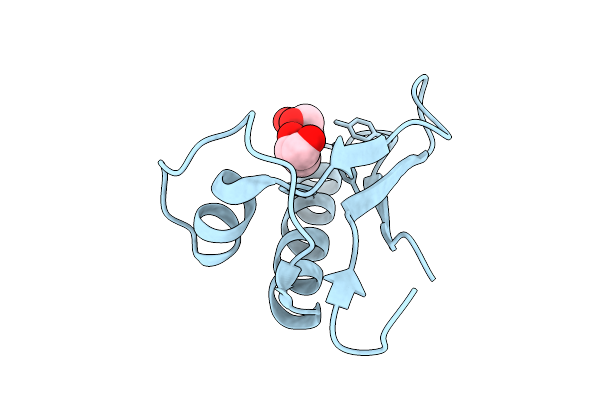 |
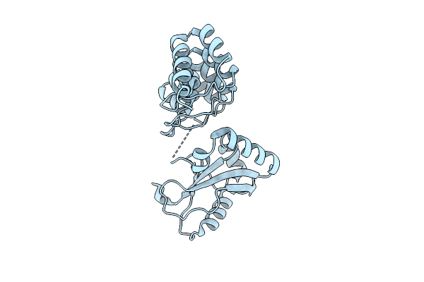
The Crystal Structure Of Nuclease Encoded By Gene Paop5_157 From Phage Paop5
Organism: Pseudomonas phage paop5
Method: X-RAY DIFFRACTION Release Date: 2025-12-31 Classification: VIRAL PROTEIN Ligands: CL, GOL |
 |
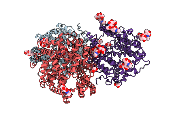
Crystal Structure Of A Putative Phosphate Binding Protein From Synechocystis Sp. Pcc 6803 Reveals An Evolutionary Hotspot
Organism: Synechocystis sp. (strain atcc 27184 / pcc 6803 / kazusa)
Method: X-RAY DIFFRACTION Release Date: 2025-12-31 Classification: PROTEIN TRANSPORT Ligands: GOL, PO4 |
 |
Organism: Caenorhabditis elegans
Method: ELECTRON MICROSCOPY Release Date: 2025-12-24 Classification: MEMBRANE PROTEIN Ligands: NAG, CLR |
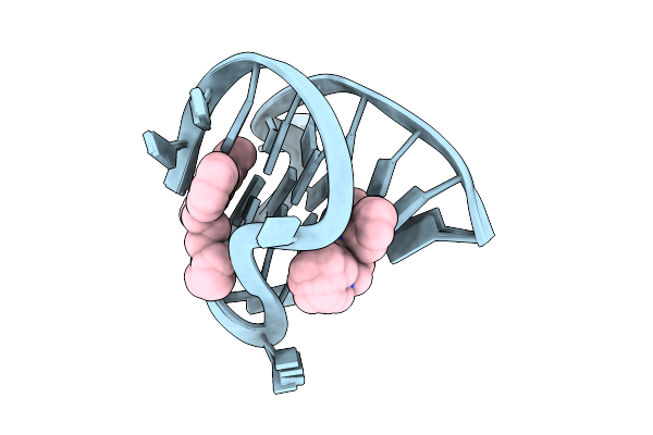 |
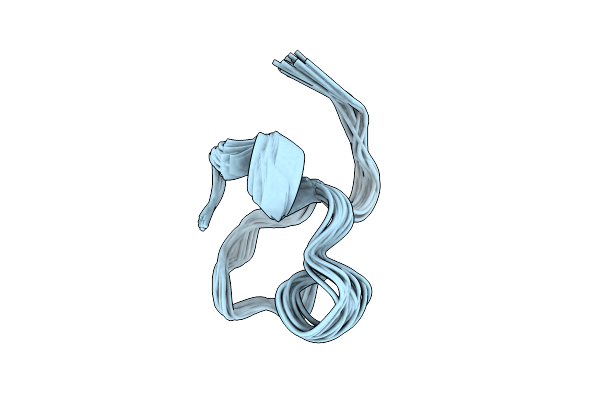
Solution Structure Of The 2:1 Complex Between The Benzothiazole Derivative Bto-28 And The Myc G-Quadruplex
Organism: Homo sapiens
Method: SOLUTION NMR Release Date: 2025-12-24 Classification: DNA Ligands: A1EIH |
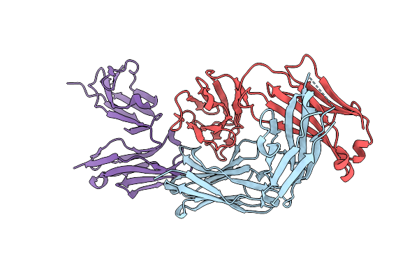 |
Organism: Homo sapiens
Method: X-RAY DIFFRACTION Release Date: 2025-12-17 Classification: IMMUNE SYSTEM |
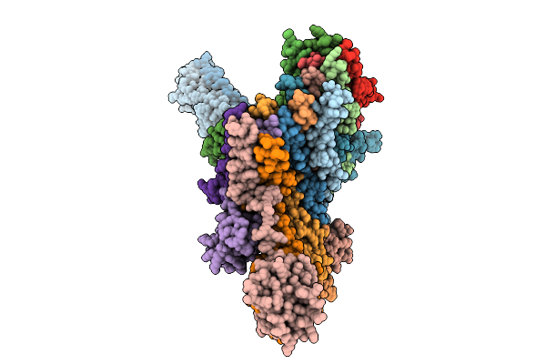 |
Cryo-Em Structure Of The Vaccinia Virus Entry/Fusion Complex (Efc) Lacking The F9 Subunit
Organism: Orthopoxvirus vaccinia
Method: ELECTRON MICROSCOPY Release Date: 2025-12-17 Classification: VIRAL PROTEIN |
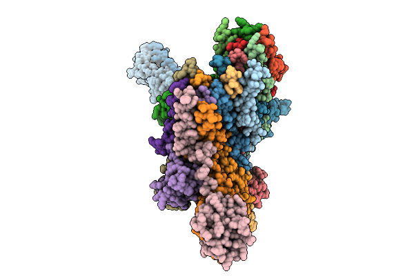 |
Cryo-Em Structure Of The Vaccinia Virus Entry/Fusion Complex (Efc) Including The F9 Subunit
Organism: Orthopoxvirus vaccinia
Method: ELECTRON MICROSCOPY Release Date: 2025-12-17 Classification: VIRAL PROTEIN |
 |
Organism: Homo sapiens
Method: SOLUTION NMR Release Date: 2025-12-10 Classification: MEMBRANE PROTEIN |
 |
Organism: Saccharomyces cerevisiae s288c
Method: SOLUTION NMR Release Date: 2025-12-10 Classification: CHAPERONE |
 |
Organism: Homo sapiens
Method: X-RAY DIFFRACTION Release Date: 2025-12-03 Classification: DNA BINDING PROTEIN |
 |
Organism: Gallus gallus
Method: X-RAY DIFFRACTION Release Date: 2025-11-26 Classification: MEMBRANE PROTEIN Ligands: NAG, EDO |
 |
Organism: Ovis aries, Bat coronavirus ratg13
Method: X-RAY DIFFRACTION Release Date: 2025-11-26 Classification: VIRAL PROTEIN/HYDROLASE Ligands: NAG |
 |
Organism: Bos taurus, Bat coronavirus ratg13
Method: X-RAY DIFFRACTION Release Date: 2025-11-26 Classification: VIRAL PROTEIN/HYDROLASE Ligands: NAG |
 |
Organism: Rattus norvegicus
Method: X-RAY DIFFRACTION Release Date: 2025-11-26 Classification: MEMBRANE PROTEIN |
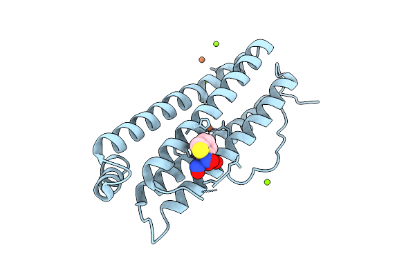 |
Crystal Structure Of Human Heavy Chain Ferritin Binding With Dinitrosyl Iron Complex Modificated By Phenylboronic Acid
Organism: Homo sapiens
Method: X-RAY DIFFRACTION Release Date: 2025-11-26 Classification: METAL BINDING PROTEIN Ligands: FE, MG, CL, NO, A1EKS, H2S |
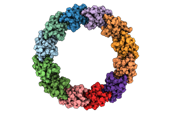 |
Organism: Escherichia phage lambda
Method: ELECTRON MICROSCOPY Release Date: 2025-11-26 Classification: VIRAL PROTEIN |
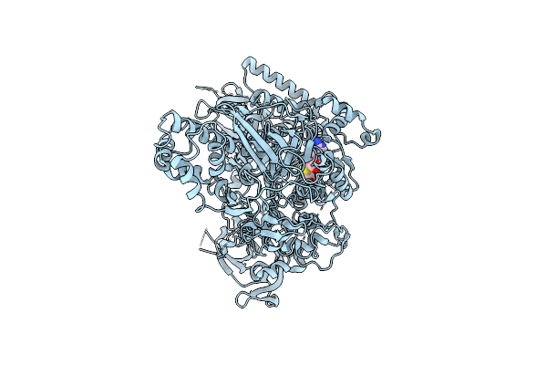 |
Organism: Arabidopsis thaliana
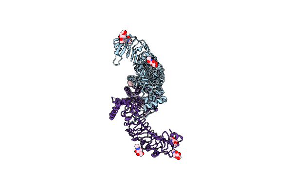
Method: ELECTRON MICROSCOPY Release Date: 2025-11-26 Classification: GENE REGULATION Ligands: SAH, ZN |
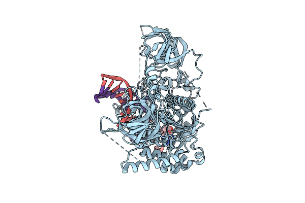 |
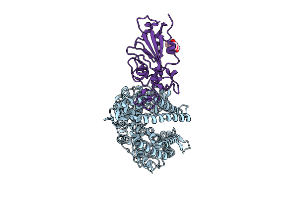
Cryo-Em Structure Of A. Thaliana Met1(610-1534) Bound Covalently To A Hemi-Mcpg Dna
Organism: Arabidopsis thaliana, Synthetic construct
Method: ELECTRON MICROSCOPY Release Date: 2025-11-26 Classification: GENE REGULATION/DNA Ligands: SAH, ZN |
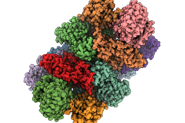 |
Organism: Homo sapiens
Method: ELECTRON MICROSCOPY Release Date: 2025-11-19 Classification: TRANSLATION |
 |
Organism: Homo sapiens
Method: ELECTRON MICROSCOPY Release Date: 2025-11-19 Classification: TRANSLATION Ligands: GTP |