
Search Count: 88
 |
Organism: Bat coronavirus
Method: ELECTRON MICROSCOPY Release Date: 2022-11-30 Classification: VIRAL PROTEIN Ligands: NAG |
 |
Organism: Pipistrellus pipistrellus, Coronavirus neoromicia/pml-phe1/rsa/2011
Method: ELECTRON MICROSCOPY Release Date: 2022-11-30 Classification: VIRAL PROTEIN Ligands: NAG |
 |
Organism: Pipistrellus pipistrellus, Bat coronavirus
Method: ELECTRON MICROSCOPY Release Date: 2022-11-30 Classification: VIRAL PROTEIN Ligands: NAG |
 |
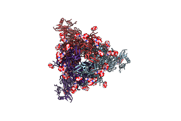
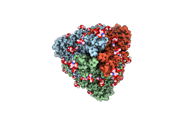
Organism: Severe acute respiratory syndrome coronavirus 2
Method: ELECTRON MICROSCOPY Release Date: 2022-11-30 Classification: VIRAL PROTEIN Ligands: NAG |
 |
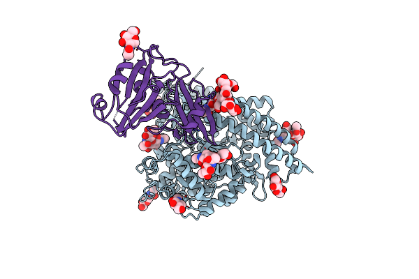
Sars-Cov-2 Ba.1 Spike Ectodomain Trimer In Complex With The S2X324 Neutralizing Antibody Fab Fragment (Local Refinement Of The Rbd And S2X324)
Organism: Homo sapiens, Severe acute respiratory syndrome coronavirus 2
Method: ELECTRON MICROSCOPY Release Date: 2022-10-26 Classification: VIRAL PROTEIN/IMMUNE SYSTEM Ligands: NAG |
 |
Sars-Cov-2 Omicron Ba.1 Spike Ectodomain Trimer In Complex With The S2X324 Neutralizing Antibody Fab Fragment
Organism: Severe acute respiratory syndrome coronavirus 2, Homo sapiens
Method: ELECTRON MICROSCOPY Release Date: 2022-10-26 Classification: VIRAL PROTEIN/IMMUNE SYSTEM Ligands: NAG |
 |
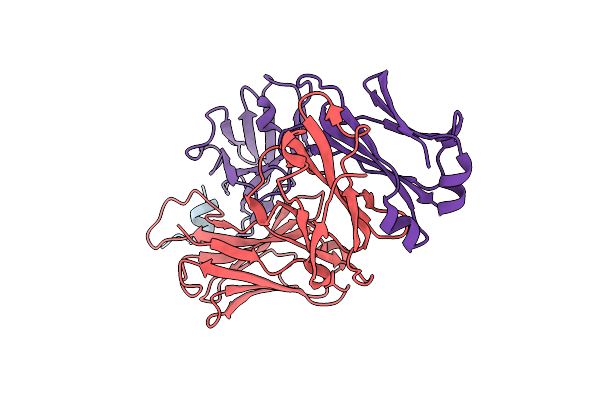
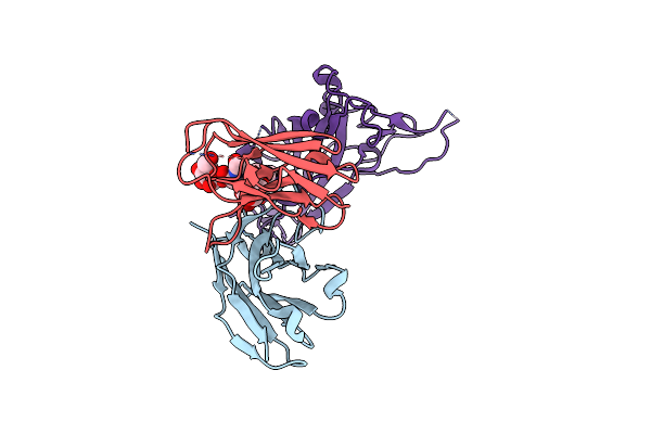
Crystal Structure Of The Ccov-Hupn-2018 Rbd (Domain B) In Complex With Canine Apn
Organism: Canis lupus familiaris, Coronaviridae sp.
Method: X-RAY DIFFRACTION Resolution:3.30 Å Release Date: 2022-08-24 Classification: VIRAL PROTEIN/IMMUNE SYSTEM Ligands: ZN, NAG |
 |
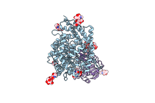
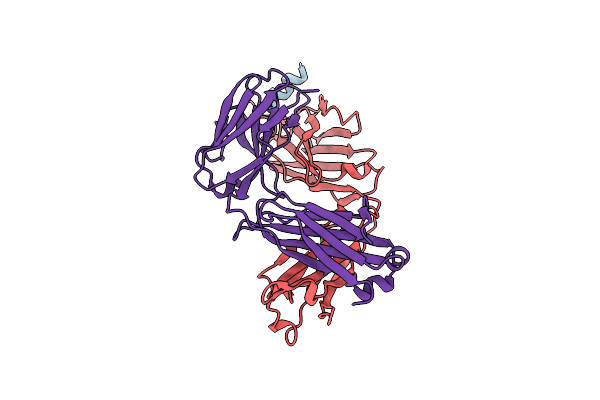
Structure Of The Human Coronavirus Ccov-Hupn-2018 Spike Glycoprotein With Domain 0 In The Proximal Conformation
Organism: Unidentified human coronavirus
Method: ELECTRON MICROSCOPY Release Date: 2022-08-24 Classification: VIRAL PROTEIN Ligands: NAG |
 |
Ccov-Hupn-2018 S In The Proximal Conformation (Local Refinement Of Domain 0)
Organism: Unidentified human coronavirus
Method: ELECTRON MICROSCOPY Release Date: 2022-08-24 Classification: VIRAL PROTEIN Ligands: NAG |
 |
Structure Of The Human Coronavirus Ccov-Hupn-2018 Spike Glycoprotein With Domain 0 In The Swung Out Conformation
Organism: Unidentified human coronavirus
Method: ELECTRON MICROSCOPY Release Date: 2022-08-24 Classification: VIRAL PROTEIN Ligands: NAG |
 |
Ccov-Hupn-2018 S In The Swung Out Conformation (Local Refinement Of Domain 0)
Organism: Unidentified human coronavirus
Method: ELECTRON MICROSCOPY Release Date: 2022-08-24 Classification: VIRAL PROTEIN Ligands: NAG |
 |
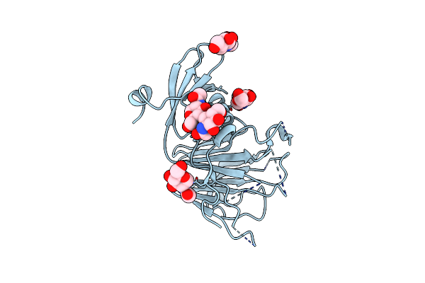
Organism: Homo sapiens, Severe acute respiratory syndrome coronavirus 2
Method: X-RAY DIFFRACTION Resolution:2.10 Å Release Date: 2022-08-03 Classification: VIRAL PROTEIN/IMMUNE SYSTEM |
 |
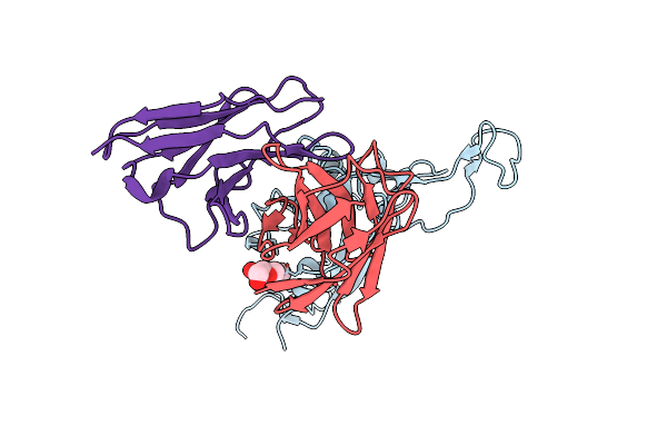
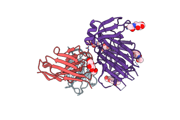
Crystal Structure Of C77G12 Fab In Complex With Sars-Cov-2 S Fusion Peptide
Organism: Homo sapiens, severe acute respiratory syndrome coronavirus 2
Method: X-RAY DIFFRACTION Resolution:1.70 Å Release Date: 2022-08-03 Classification: VIRAL PROTEIN/IMMUNE SYSTEM Ligands: CL |
 |
Organism: Homo sapiens, Severe acute respiratory syndrome coronavirus 2
Method: X-RAY DIFFRACTION Resolution:2.10 Å Release Date: 2022-08-03 Classification: VIRAL PROTEIN/IMMUNE SYSTEM |
 |
Crystal Structure Of Vn01H1 Fab In Complex With Sars-Cov-2 S Fusion Peptide
Organism: Homo sapiens, Severe acute respiratory syndrome coronavirus 2
Method: X-RAY DIFFRACTION Resolution:1.86 Å Release Date: 2022-07-20 Classification: VIRAL PROTEIN/IMMUNE SYSTEM |
 |
Crystal Structure Of Vp12E7 Fab In Complex With Sars-Cov-2 S Fusion Peptide
Organism: Homo sapiens, Severe acute respiratory syndrome coronavirus 2
Method: X-RAY DIFFRACTION Resolution:2.50 Å Release Date: 2022-07-20 Classification: VIRAL PROTEIN/IMMUNE SYSTEM |
 |
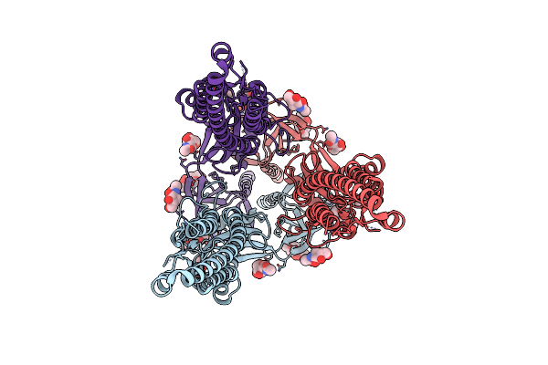
Sars-Cov-2 Spike In Complex With Ahb2-2Gs-Sb175 (Local Refinement Of The Rbd And Ahb2)
Organism: Severe acute respiratory syndrome coronavirus, Synthetic construct
Method: ELECTRON MICROSCOPY Release Date: 2022-06-08 Classification: VIRAL PROTEIN |
 |
Organism: Severe acute respiratory syndrome coronavirus 2, Synthetic construct
Method: ELECTRON MICROSCOPY Release Date: 2022-06-08 Classification: VIRAL PROTEIN Ligands: NAG |
 |
Organism: Homo sapiens, Severe acute respiratory syndrome coronavirus 2
Method: ELECTRON MICROSCOPY Release Date: 2022-02-02 Classification: VIRUS/IMMUNE SYSTEM |
 |
Organism: Homo sapiens
Method: ELECTRON MICROSCOPY Release Date: 2022-02-02 Classification: VIRUS/IMMUNE SYSTEM Ligands: NAG |