
Search Count: 14
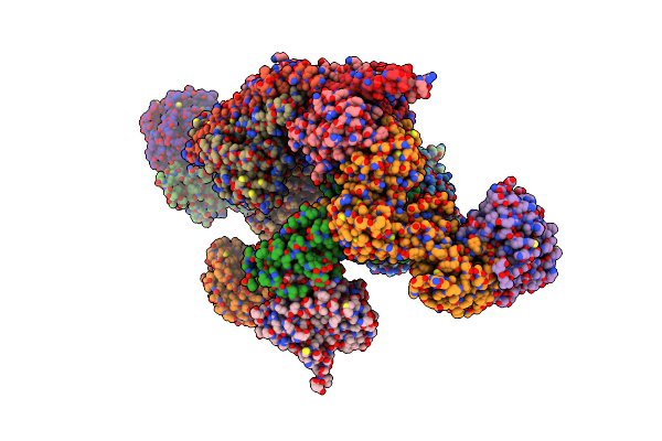 |
Cryo-Em Structure Of The Human Brisc Dimer Complex Bound To Compound Fx-171-C
Organism: Homo sapiens
Method: ELECTRON MICROSCOPY Release Date: 2025-02-12 Classification: SIGNALING PROTEIN Ligands: ZN, G1V |
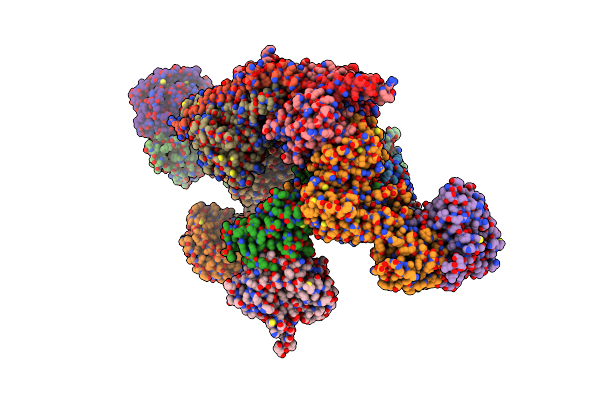 |
Cryo-Em Structure Of The Human Brisc Dimer Complex Bound To Compound Jms-175-2
Organism: Homo sapiens
Method: ELECTRON MICROSCOPY Release Date: 2025-02-05 Classification: SIGNALING PROTEIN Ligands: ZN, X8C |
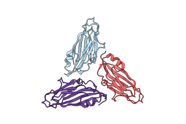 |
Organism: Barley yellow dwarf virus
Method: ELECTRON MICROSCOPY Release Date: 2019-10-02 Classification: VIRUS LIKE PARTICLE |
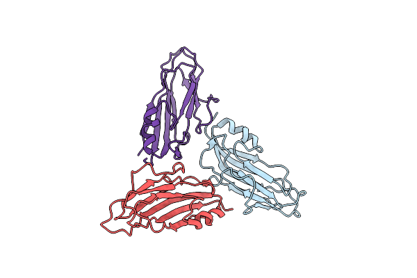 |
Organism: Potato leafroll virus
Method: ELECTRON MICROSCOPY Release Date: 2019-10-02 Classification: VIRUS LIKE PARTICLE |
 |
Organism: Homo sapiens
Method: ELECTRON MICROSCOPY Release Date: 2019-06-05 Classification: SIGNALING PROTEIN Ligands: ZN |
 |
N-Terminal Thioester Domain Of Surface Protein From Clostridium Perfringens, Cys138Ala Mutant
Organism: Clostridium perfringens
Method: X-RAY DIFFRACTION Resolution:1.60 Å Release Date: 2015-06-03 Classification: CELL ADHESION Ligands: EDO |
 |
N-Terminal Thioester Domain Of Surface Protein From Clostridium Perfringens
Organism: Clostridium perfringens
Method: X-RAY DIFFRACTION Resolution:2.62 Å Release Date: 2015-06-03 Classification: CELL ADHESION |
 |
N-Terminal Thioester Domain Of Fibronectin-Binding Protein Sfbi From Streptococcus Pyogenes
Organism: Streptococcus pyogenes
Method: X-RAY DIFFRACTION Resolution:1.35 Å Release Date: 2015-06-03 Classification: CELL ADHESION Ligands: ZN, ACT |
 |
N-Terminal Thioester Domain Of Protein F2 Like Fibronectin-Binding Protein From Streptococcus Pneumoniae
Organism: Streptococcus pneumoniae
Method: X-RAY DIFFRACTION Resolution:1.30 Å Release Date: 2015-06-03 Classification: CELL ADHESION Ligands: MES, GOL |
 |
Organism: Streptococcus pyogenes
Method: X-RAY DIFFRACTION Resolution:2.80 Å Release Date: 2013-10-09 Classification: CELL ADHESION |
 |
Organism: Drosophila melanogaster
Method: X-RAY DIFFRACTION Resolution:1.39 Å Release Date: 2011-07-06 Classification: HYDROLASE Ligands: SO4, GNP, MG |
 |
Organism: Escherichia coli, Homo sapiens
Method: X-RAY DIFFRACTION Resolution:3.97 Å Release Date: 2006-01-03 Classification: PROTON TRANSPORT,MEMBRANE PROTEIN |
 |
Organism: Escherichia coli, Homo sapiens
Method: X-RAY DIFFRACTION Resolution:3.32 Å Release Date: 2006-01-03 Classification: PROTON TRANSPORT,MEMBRANE PROTEIN |
 |
Organism: Escherichia coli, Homo sapiens
Method: X-RAY DIFFRACTION Resolution:3.20 Å Release Date: 2006-01-03 Classification: PROTON TRANSPORT,MEMBRANE PROTEIN |