
Search Count: 21
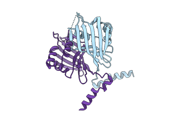 |
Organism: Mus musculus
Method: X-RAY DIFFRACTION Release Date: 2025-08-13 Classification: STRUCTURAL PROTEIN |
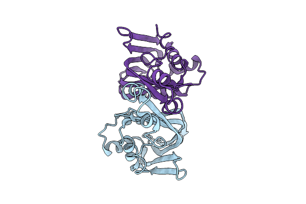 |
Organism: Mus musculus
Method: X-RAY DIFFRACTION Release Date: 2025-07-30 Classification: STRUCTURAL PROTEIN |
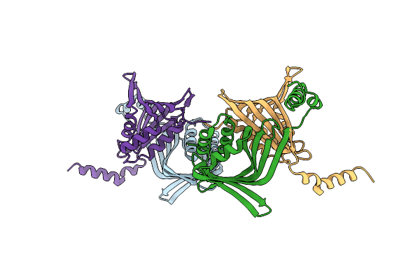 |
Organism: Mus musculus
Method: X-RAY DIFFRACTION Resolution:2.30 Å Release Date: 2025-03-19 Classification: GENE REGULATION |
 |
E. Coli 70S Ribosome Complexed With P. Putida Trnaile2 At The A-Site And P-Site
Organism: Escherichia coli, Pseudomonas putida nbrc 14164
Method: ELECTRON MICROSCOPY Release Date: 2024-11-06 Classification: RIBOSOME Ligands: MG |
 |
Organism: Escherichia coli, Escherichia coli bw25113, Pseudomonas putida nbrc 14164
Method: ELECTRON MICROSCOPY Release Date: 2024-11-06 Classification: RIBOSOME Ligands: MG |
 |
Organism: Escherichia coli, Escherichia coli bw25113, Pseudomonas putida nbrc 14164
Method: ELECTRON MICROSCOPY Release Date: 2024-11-06 Classification: RIBOSOME Ligands: MG |
 |
Organism: Escherichia coli, Escherichia coli bw25113, Pseudomonas putida nbrc 14164
Method: ELECTRON MICROSCOPY Release Date: 2024-11-06 Classification: RIBOSOME Ligands: MG |
 |
Organism: Escherichia coli, Escherichia coli bw25113, Pseudomonas putida nbrc 14164
Method: ELECTRON MICROSCOPY Release Date: 2024-11-06 Classification: RIBOSOME Ligands: MG |
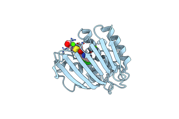 |
Pseudomonas Aeruginosa Dna Gyrase B 24Kda Atpase Subdomain Complexed With Ebl3021
Organism: Pseudomonas aeruginosa pao1
Method: X-RAY DIFFRACTION Resolution:1.60 Å Release Date: 2023-03-29 Classification: DNA BINDING PROTEIN Ligands: CA, R53 |
 |
Organism: Lactococcus lactis
Method: X-RAY DIFFRACTION Resolution:3.10 Å Release Date: 2017-08-16 Classification: LIGASE Ligands: MN, BTN |
 |
Crystal Structure Of Lactococcus Lactis Pyruvate Carboxylase In Complex With Cyclic-Di-Amp
Organism: Lactococcus lactis
Method: X-RAY DIFFRACTION Resolution:2.30 Å Release Date: 2017-08-16 Classification: LIGASE Ligands: MN, ADP, MG, 2BA |
 |
Crystal Structure Of Lactococcus Lactis Pyruvate Carboxylase G746A Mutant In Complex With Cyclic-Di-Amp
Organism: Lactococcus lactis
Method: X-RAY DIFFRACTION Resolution:2.00 Å Release Date: 2017-08-16 Classification: LIGASE Ligands: MN, ADP, MG, 2BA |
 |
Organism: Neurospora crassa
Method: X-RAY DIFFRACTION Resolution:2.10 Å Release Date: 2008-11-11 Classification: TRANSFERASE Ligands: P6G |
 |
X-Ray Crystal Structure Of A Type Iii Pentaketide Synthase From Neurospora Crassa
Organism: Neurospora crassa
Method: X-RAY DIFFRACTION Resolution:2.00 Å Release Date: 2008-11-11 Classification: TRANSFERASE Ligands: DCR |
 |
Organism: Neurospora crassa
Method: X-RAY DIFFRACTION Resolution:1.75 Å Release Date: 2008-11-04 Classification: TRANSFERASE |
 |
Crystal Structure Of The Aromatase/Cyclase Domain Of Tcmn From Streptomyces Glaucescens
Organism: Streptomyces glaucescens
Method: X-RAY DIFFRACTION Resolution:1.90 Å Release Date: 2008-04-22 Classification: BIOSYNTHETIC PROTEIN |
 |
Organism: Streptomyces glaucescens
Method: X-RAY DIFFRACTION Resolution:2.20 Å Release Date: 2008-04-22 Classification: BIOSYNTHETIC PROTEIN |
 |
Organism: Streptomyces glaucescens
Method: X-RAY DIFFRACTION Resolution:1.95 Å Release Date: 2008-04-22 Classification: BIOSYNTHETIC PROTEIN Ligands: ACT, IOD |
 |
Actinorhodin Polyketide Ketoreductase With Nadp And The Inhibitor Isoniazid Bound
Organism: Streptomyces coelicolor
Method: X-RAY DIFFRACTION Resolution:2.71 Å Release Date: 2005-09-27 Classification: OXIDOREDUCTASE Ligands: NAP, ISZ |
 |
Organism: Streptomyces coelicolor
Method: X-RAY DIFFRACTION Resolution:2.30 Å Release Date: 2004-12-14 Classification: OXIDOREDUCTASE Ligands: NAP |