
Search Count: 37
 |
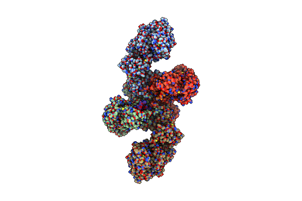
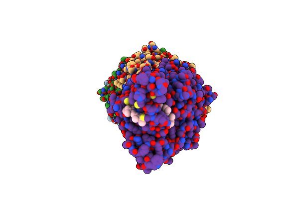
Structure Of The Non-Canonical Ctlh E3 Substrate Receptor Wdr26 Bound To Ypel5
Organism: Homo sapiens
Method: ELECTRON MICROSCOPY Release Date: 2024-05-15 Classification: LIGASE Ligands: ZN |
 |
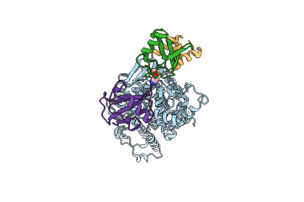
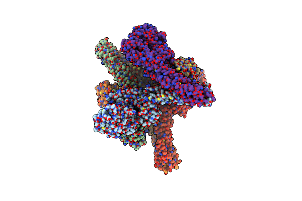
Structure Of The Non-Canonical Ctlh E3 Substrate Receptor Wdr26 Bound To Nmnat1 Substrate
Organism: Homo sapiens
Method: ELECTRON MICROSCOPY Release Date: 2024-05-15 Classification: LIGASE Ligands: NMN, ZN |
 |
 |
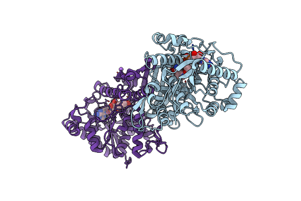
Structure Of Cul9-Rbx1 Ubiquitin E3 Ligase Complex In Unneddylated And Neddylated Conformation - Focused Cullin Dimer
Organism: Homo sapiens
Method: ELECTRON MICROSCOPY Release Date: 2024-04-17 Classification: LIGASE Ligands: ZN |
 |
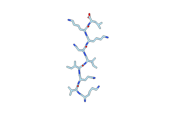
Structure Of Cul9-Rbx1 Ubiquitin E3 Ligase Complex In Unneddylated Conformation - Symmetry Expanded Unneddylated Dimer
Organism: Homo sapiens
Method: ELECTRON MICROSCOPY Release Date: 2024-04-17 Classification: LIGASE Ligands: ZN |
 |
Catalytic Module Of Human Ctlh E3 Ligase Bound To Multiphosphorylated Ube2H~Ubiquitin
Organism: Homo sapiens
Method: ELECTRON MICROSCOPY Release Date: 2024-01-03 Classification: LIGASE Ligands: ZN |
 |
Catalytic Module Of Yeast Gid E3 Ligase Bound To Multiphosphorylated Ubc8~Ubiquitin
Organism: Saccharomyces cerevisiae, Homo sapiens
Method: ELECTRON MICROSCOPY Release Date: 2024-01-03 Classification: LIGASE Ligands: ZN |
 |
Organism: Homo sapiens
Method: ELECTRON MICROSCOPY Release Date: 2023-08-23 Classification: LIGASE Ligands: ZN |
 |
Organism: Homo sapiens
Method: ELECTRON MICROSCOPY Release Date: 2023-08-23 Classification: LIGASE Ligands: SY8 |
 |
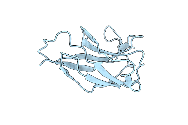
Crystal Structure Of The Rabies Virus Rna Free Nucleoprotein- Phosphoprotein Complex
Organism: Lyssavirus rabies
Method: X-RAY DIFFRACTION Resolution:2.30 Å Release Date: 2023-03-15 Classification: VIRAL PROTEIN Ligands: PEG |
 |
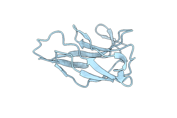
Organism: Rabies virus cvs-11, Synthetic construct
Method: X-RAY DIFFRACTION Resolution:3.49 Å Release Date: 2023-01-11 Classification: VIRAL PROTEIN Ligands: PO4 |
 |
Organism: Saccharomyces cerevisiae yjm1133, Saccharomyces cerevisiae
Method: ELECTRON MICROSCOPY Release Date: 2022-06-08 Classification: LIGASE |
 |
Organism: Bacteroides thetaiotaomicron
Method: X-RAY DIFFRACTION Resolution:2.00 Å Release Date: 2022-03-30 Classification: ISOMERASE Ligands: NAD, NA |
 |
Organism: Homo sapiens
Method: ELECTRON CRYSTALLOGRAPHY Resolution:1.99 Å Release Date: 2020-01-15 Classification: STRUCTURAL PROTEIN |
 |
Organism: Geobacter sulfurreducens (strain atcc 51573 / dsm 12127 / pca)
Method: ELECTRON MICROSCOPY Release Date: 2019-04-10 Classification: ELECTRON TRANSPORT Ligands: HEC |
 |
Organism: Mus musculus
Method: X-RAY DIFFRACTION Resolution:3.00 Å Release Date: 2014-01-15 Classification: TRANSCRIPTION |
 |
Organism: Mus musculus
Method: X-RAY DIFFRACTION Resolution:2.44 Å Release Date: 2013-09-04 Classification: TRANSCRIPTION |
 |
Organism: Homo sapiens
Method: X-RAY DIFFRACTION Resolution:3.98 Å Release Date: 2013-06-19 Classification: TRANSFERASE |
 |
Organism: Mus musculus
Method: X-RAY DIFFRACTION Resolution:2.60 Å Release Date: 2010-11-24 Classification: TRANSCRIPTION |
 |
Organism: Mus musculus
Method: X-RAY DIFFRACTION Resolution:2.51 Å Release Date: 2010-11-24 Classification: TRANSCRIPTION |