
Search Count: 21
 |
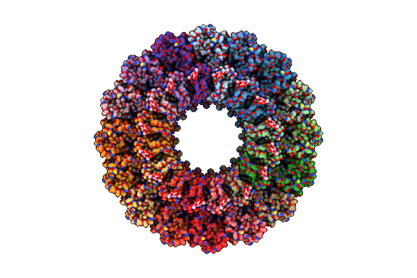
Organism: Mus musculus
Method: ELECTRON MICROSCOPY Release Date: 2022-02-16 Classification: IMMUNE SYSTEM Ligands: CA, NAG |
 |
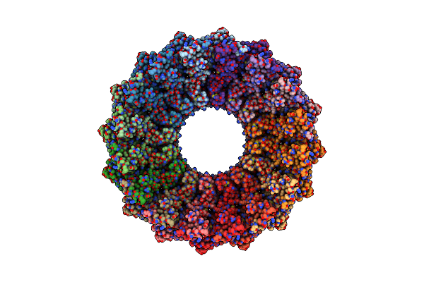
Organism: Homo sapiens
Method: ELECTRON MICROSCOPY Release Date: 2019-09-25 Classification: IMMUNE SYSTEM |
 |
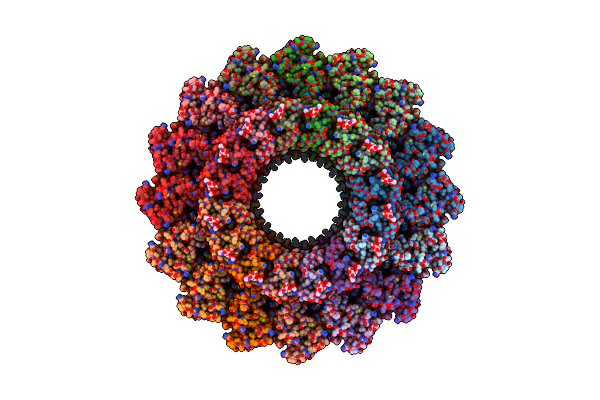
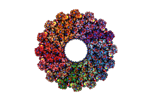
Em Structure Of Mpeg-1 (L425K, Alpha Conformation) Soluble Pre-Pore Complex
Organism: Homo sapiens
Method: ELECTRON MICROSCOPY Release Date: 2019-09-25 Classification: IMMUNE SYSTEM Ligands: NAG |
 |
Em Structure Of Mpeg-1 (L425K, Alpha Conformation) Soluble Pre-Pore Complex
Organism: Homo sapiens
Method: ELECTRON MICROSCOPY Release Date: 2019-09-25 Classification: IMMUNE SYSTEM |
 |
Organism: Homo sapiens
Method: ELECTRON MICROSCOPY Release Date: 2019-09-25 Classification: IMMUNE SYSTEM Ligands: NAG |
 |
Organism: Homo sapiens
Method: ELECTRON MICROSCOPY Release Date: 2019-09-25 Classification: IMMUNE SYSTEM |
 |
Organism: Mus musculus
Method: X-RAY DIFFRACTION Resolution:2.00 Å Release Date: 2018-02-07 Classification: APOPTOSIS Ligands: IOD |
 |
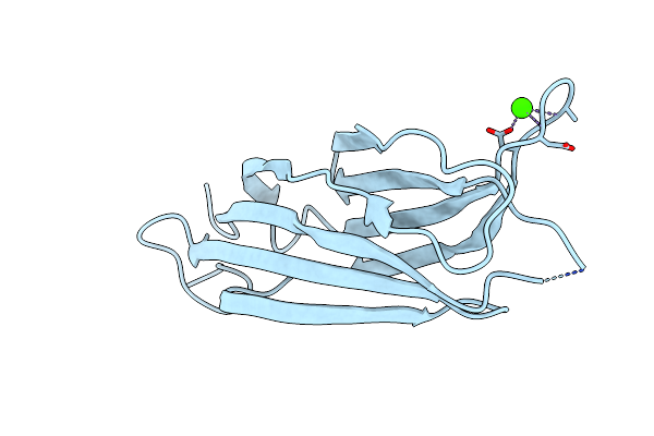
Organism: Mus musculus
Method: X-RAY DIFFRACTION Resolution:1.80 Å Release Date: 2018-02-07 Classification: APOPTOSIS Ligands: CA |
 |
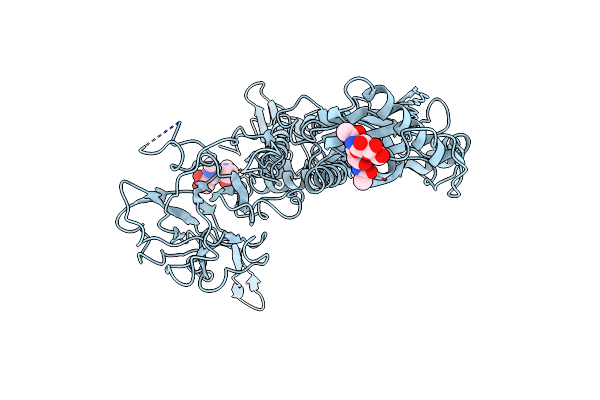
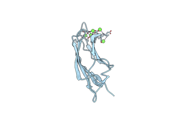
Structural Basis For Ca2+-Mediated Interaction Of The Perforin C2 Domain With Lipid Membranes
Organism: Mus musculus
Method: X-RAY DIFFRACTION Resolution:1.61 Å Release Date: 2015-09-02 Classification: IMMUNE SYSTEM Ligands: CA |
 |
Structural Basis For Ca2+-Mediated Interaction Of The Perforin C2 Domain With Lipid Membranes
Organism: Mus musculus
Method: X-RAY DIFFRACTION Resolution:2.67 Å Release Date: 2015-09-02 Classification: IMMUNE SYSTEM Ligands: CA |
 |
Structure Of Membrane Binding Protein Pleurotolysin A From Pleurotus Ostreatus
Organism: Pleurotus ostreatus
Method: X-RAY DIFFRACTION Resolution:1.85 Å Release Date: 2015-02-18 Classification: MEMBRANE BINDING PROTEIN Ligands: SO4 |
 |
Structure Of Membrane Binding Protein Pleurotolysin B From Pleurotus Ostreatus
Organism: Pleurotus ostreatus
Method: X-RAY DIFFRACTION Resolution:2.20 Å Release Date: 2015-02-18 Classification: MEMBRANE BINDING PROTEIN Ligands: ACT, GOL, CL |
 |
Crystal Structure Of The Tmh1-Lock Mutant Of The Mature Form Of Pleurotolysin B
Organism: Pleurotus ostreatus
Method: X-RAY DIFFRACTION Resolution:2.15 Å Release Date: 2015-02-18 Classification: TOXIN Ligands: SO4, CL, GOL |
 |
Organism: Pleurotus ostreatus
Method: ELECTRON MICROSCOPY Resolution:11.00 Å Release Date: 2015-02-18 Classification: TRANSPORT PROTEIN |
 |
Membrane Bound Pleurotolysin Prepore (Tmh1 Lock) Trapped With Engineered Disulphide Cross-Link
Organism: Pleurotus ostreatus
Method: ELECTRON MICROSCOPY Resolution:15.00 Å Release Date: 2015-02-18 Classification: TRANSPORT PROTEIN |
 |
Membrane Bound Pleurotolysin Prepore (Tmh2 Helix Lock) Trapped With Engineered Disulphide Cross-Link
Organism: Pleurotus ostreatus
Method: ELECTRON MICROSCOPY Resolution:17.00 Å Release Date: 2015-02-18 Classification: TRANSPORT PROTEIN |
 |
Membrane Bound Pleurotolysin Prepore (Tmh2 Strand Lock) Trapped With Engineered Disulphide Cross-Link
Organism: Pleurotus ostreatus
Method: ELECTRON MICROSCOPY Resolution:14.00 Å Release Date: 2015-02-18 Classification: TRANSPORT PROTEIN |
 |
Organism: Scophthalmus maximus
Method: X-RAY DIFFRACTION Resolution:1.60 Å Release Date: 2013-10-23 Classification: LIPID BINDING PROTEIN |
 |
Organism: Scophthalmus maximus
Method: X-RAY DIFFRACTION Resolution:1.66 Å Release Date: 2013-10-23 Classification: LIPID BINDING PROTEIN Ligands: CA |
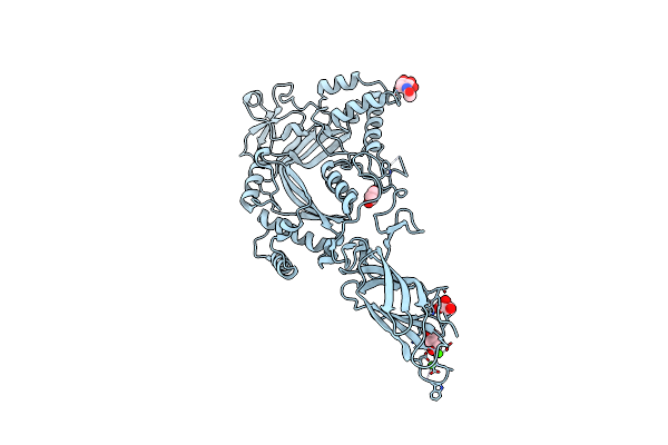 |
Organism: Mus musculus
Method: X-RAY DIFFRACTION Resolution:2.75 Å Release Date: 2010-11-03 Classification: IMMUNE SYSTEM Ligands: GOL, CA, IOD, CL, NAG |