
Search Count: 264
 |
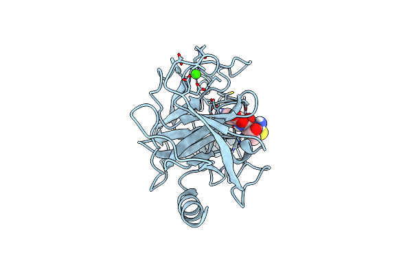
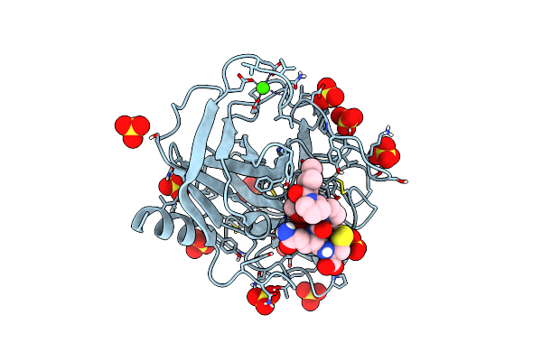
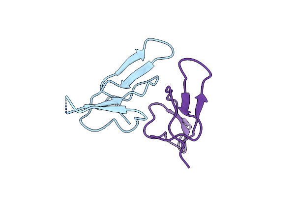
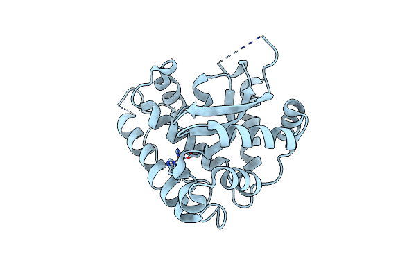
Crystallographic Structure Of The Octapeptide Derived From The Btci Inhibitor Bound To Beta-Trypsin In Space Group P 21 21 21.
Organism: Vigna unguiculata, Bos taurus
Method: X-RAY DIFFRACTION Resolution:1.18 Å Release Date: 2019-08-07 Classification: hydrolase/hydrolase inhibitor Ligands: SO4, CA |
 |
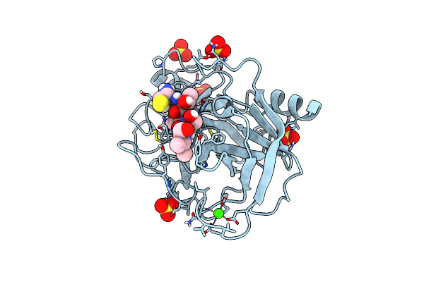
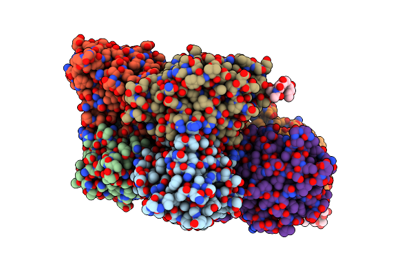
Crystallographic Structure Of The Cyclic Heptapeptide Derived From The Btci Inhibitor Bound To Beta-Trypsin In Space Group P 4(1) 2(1) 2
Organism: Vigna unguiculata, Bos taurus
Method: X-RAY DIFFRACTION Resolution:1.39 Å Release Date: 2019-08-07 Classification: hydrolase/hydrolase inhibitor Ligands: CA |
 |
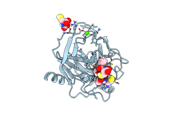
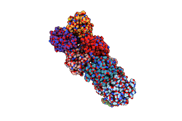
Crystallographic Structure Of The Cyclic Heptapeptide Derived From The Btci Inhibitor Bound To Beta-Trypsin In Space Group P 21 21 21
Organism: Vigna unguiculata, Bos taurus
Method: X-RAY DIFFRACTION Resolution:1.29 Å Release Date: 2019-08-07 Classification: hydrolase/hydrolase inhibitor Ligands: SO4, CA |
 |
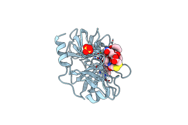
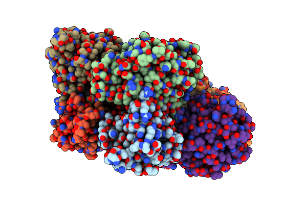
Crystallographic Structure Of The Cyclic Hexapeptide Derived From The Btci Inhibitor Bound To Beta-Trypsin In Space Group P 21 21 21
Organism: Vigna unguiculata, Bos taurus
Method: X-RAY DIFFRACTION Resolution:1.19 Å Release Date: 2019-08-07 Classification: hydrolase/hydrolase inhibitor Ligands: CA, J3D |
 |
Crystallographic Structure Of The Cyclic Nonapeptide Derived From The Btci Inhibitor Bound To Beta-Trypsin In Space Group P 32 2 1
Organism: Vigna unguiculata, Bos taurus
Method: X-RAY DIFFRACTION Resolution:1.61 Å Release Date: 2019-03-13 Classification: hydrolase/hydrolase inhibitor Ligands: CA, SO4 |
 |
Crystallographic Structure Of The Cyclic Nonapeptide Derived From The Btci Inhibitor Bound To Beta-Trypsin In Space Group P 21 21 21.
Organism: Vigna unguiculata, Bos taurus
Method: X-RAY DIFFRACTION Resolution:1.15 Å Release Date: 2019-03-13 Classification: hydrolase/hydrolase inhibitor Ligands: CA, SO4 |
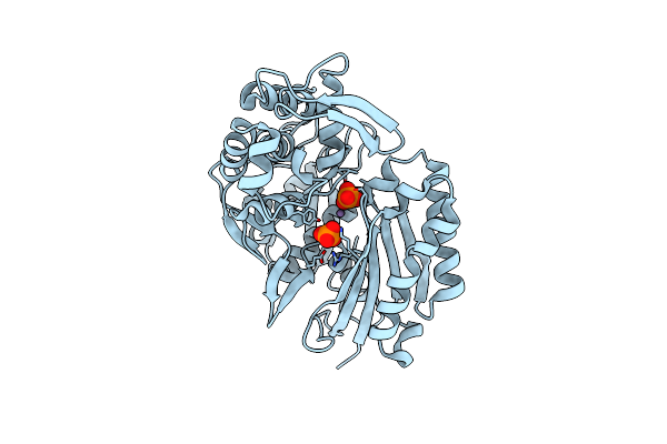 |
Crystal Structure Of A Nucleoside Triphosphate Diphosphohydrolase (Ntpdase) From The Legume Vigna Unguiculata Subsp. Cylindrica (Dolichos Biflorus) In Complex With Phosphate And Manganese
Organism: Vigna unguiculata subsp. cylindrica
Method: X-RAY DIFFRACTION Resolution:2.60 Å Release Date: 2017-05-31 Classification: HYDROLASE Ligands: PO4, MN |
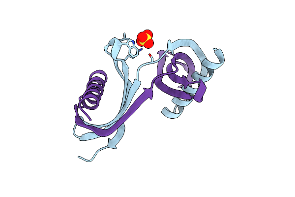 |
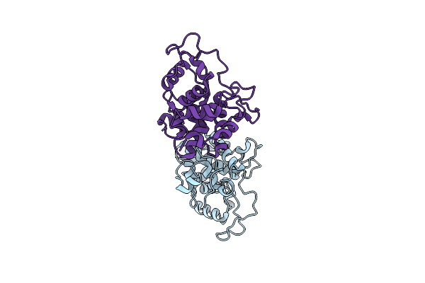
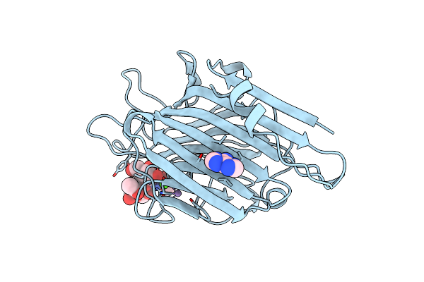
Crystal Structure Of A Single-Domain Cysteine Protease Inhibitor From Cowpea (Vigna Unguiculata)
Organism: Vigna unguiculata
Method: X-RAY DIFFRACTION Resolution:1.95 Å Release Date: 2015-10-14 Classification: HYDROLASE INHIBITOR Ligands: SO4 |
 |
Organism: Vigna unguiculata subsp. sesquipedalis
Method: X-RAY DIFFRACTION Resolution:1.55 Å Release Date: 2015-10-14 Classification: HYDROLASE |
 |
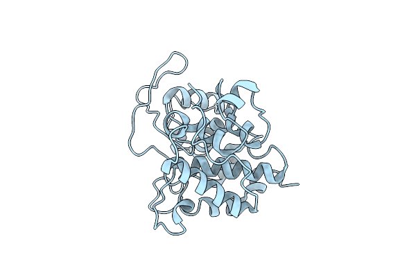
Structure Of Vigna Unguiculata Chitinase With Regulation Activity Of The Plant Cell Wall
Organism: Vigna unguiculata
Method: X-RAY DIFFRACTION Resolution:1.50 Å Release Date: 2013-01-16 Classification: HYDROLASE |
 |
Crystal Structure Of The Bowman-Birk Serine Protease Inhibitor Btci In Complex With Trypsin And Chymotrypsin
Organism: Bos taurus, Vigna unguiculata
Method: X-RAY DIFFRACTION Resolution:1.68 Å Release Date: 2012-08-29 Classification: HYDROLASE/HYDROLASE INHIBITOR Ligands: CA, GOL, SO4, EDO, MRD |
 |
Organism: Vigna unguiculata
Method: X-RAY DIFFRACTION Resolution:2.10 Å Release Date: 2011-02-09 Classification: PLANT PROTEIN Ligands: CA, CL, NA |
 |
Organism: Vigna unguiculata
Method: X-RAY DIFFRACTION Resolution:2.50 Å Release Date: 2007-11-27 Classification: PLANT PROTEIN |
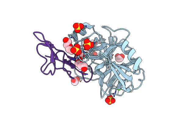 |
Crystal Structure Of The Bowman-Birk Inhibitor From Vigna Unguiculata Seeds In Complex With Beta-Trypsin At 1.55 Angstrons Resolution
Organism: Bos taurus, Vigna unguiculata
Method: X-RAY DIFFRACTION Resolution:1.55 Å Release Date: 2007-01-02 Classification: HYDROLASE/HYDROLASE INHIBITOR Ligands: CA, SO4, PGE, EDO, ACY, P6G |
 |
The Crystal Structure Of The Eukaryotic Fesod From Vigna Unguiculata Suggests A New Enzymatic Mechanism
Organism: Vigna unguiculata
Method: X-RAY DIFFRACTION Resolution:1.97 Å Release Date: 2004-10-27 Classification: OXIDOREDUCTASE Ligands: FE |
 |
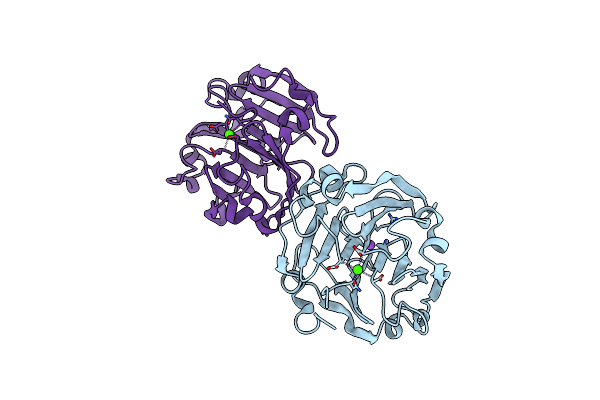
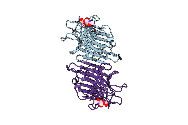
The Crystal Structure Of The 58Kd Vegetative Lectin From The Tropical Legume Dolichos Biflorus
Organism: Vigna unguiculata subsp. cylindrica
Method: X-RAY DIFFRACTION Resolution:2.50 Å Release Date: 2000-11-29 Classification: SUGAR BINDING PROTEIN Ligands: CA, MN |
 |
Organism: Vigna unguiculata subsp. cylindrica
Method: X-RAY DIFFRACTION Resolution:2.65 Å Release Date: 1998-12-30 Classification: GLYCOPROTEIN Ligands: CA, MN, ADE |
 |
Organism: Vigna unguiculata subsp. cylindrica
Method: X-RAY DIFFRACTION Resolution:3.30 Å Release Date: 1998-12-30 Classification: LECTIN Ligands: MN, CA |
 |
The Structure Of The Dolichos Biflorus Seed Lectin In Complex With The Forssman Disaccharide
Organism: Vigna unguiculata subsp. cylindrica
Method: X-RAY DIFFRACTION Resolution:2.60 Å Release Date: 1998-12-09 Classification: LECTIN Ligands: CA, MN, ADE |
 |
Dolichos Biflorus Seed Lectin In Complex With The Blood Group A Trisaccharide
Organism: Vigna unguiculata subsp. cylindrica
Method: X-RAY DIFFRACTION Resolution:2.80 Å Release Date: 1998-12-09 Classification: LECTIN Ligands: A2G, CA, MN |