
Search Count: 20
All
Selected
 |
Organism: Homo sapiens
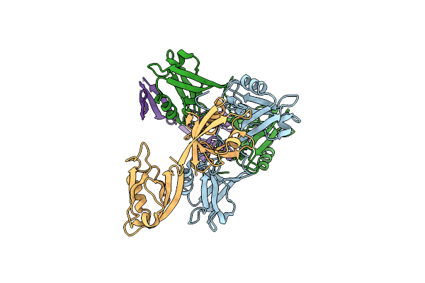
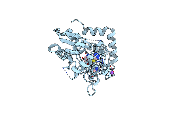
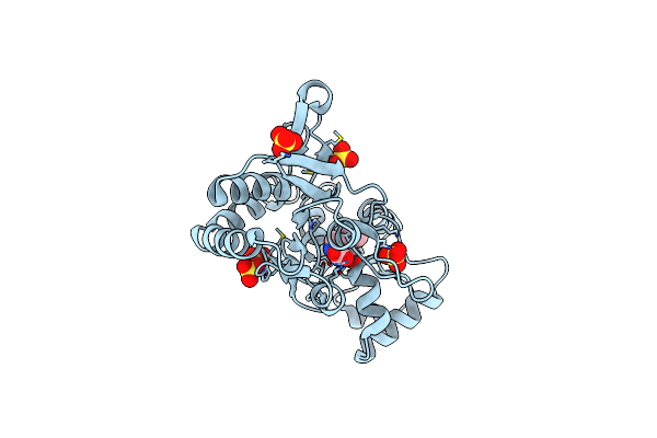
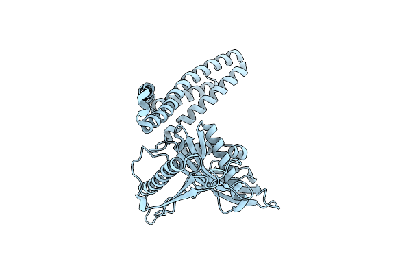
Method: X-RAY DIFFRACTION Resolution:3.40 Å Release Date: 2012-03-21 Classification: SIGNALING PROTEIN |
 |
Organism: Homo sapiens
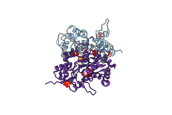
Method: X-RAY DIFFRACTION Resolution:1.98 Å Release Date: 2011-02-23 Classification: CELL ADHESION Ligands: NAG |
 |
Organism: Homo sapiens
Method: X-RAY DIFFRACTION Resolution:1.82 Å Release Date: 2011-02-23 Classification: CELL ADHESION Ligands: NAG, GOL |
 |
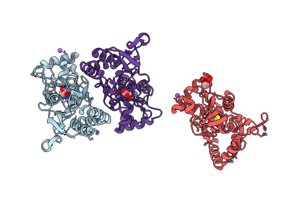
Organism: Homo sapiens
Method: X-RAY DIFFRACTION Resolution:2.51 Å Release Date: 2011-02-23 Classification: CELL ADHESION Ligands: NAG, EPE, PO4 |
 |
Organism: Homo sapiens
Method: X-RAY DIFFRACTION Resolution:3.00 Å Release Date: 2010-08-25 Classification: CELL ADHESION Ligands: NAG, CA |
 |
Organism: Homo sapiens
Method: X-RAY DIFFRACTION Resolution:2.30 Å Release Date: 2009-11-17 Classification: CELL ADHESION Ligands: NAG, GOL, CA |
 |
Structure Of The Ligand-Binding Core Of Glur2 In Complex With The Agonist (S)-Tdpa At 2.25 A Resolution
Organism: Rattus norvegicus
Method: X-RAY DIFFRACTION Resolution:2.27 Å Release Date: 2008-10-28 Classification: MEMBRANE PROTEIN Ligands: S2P, ZN, NA, CL, CAC |
 |
Structure Of The Ligand-Binding Core Of Glur2 In Complex With The Agonist (R)-Tdpa At 1.95 A Resolution
Organism: Rattus norvegicus
Method: X-RAY DIFFRACTION Resolution:1.95 Å Release Date: 2008-10-14 Classification: MEMBRANE PROTEIN Ligands: R2P |
 |
Organism: Homo sapiens
Method: X-RAY DIFFRACTION Resolution:2.00 Å Release Date: 2008-07-29 Classification: CELL ADHESION Ligands: SO4, NAG, GOL |
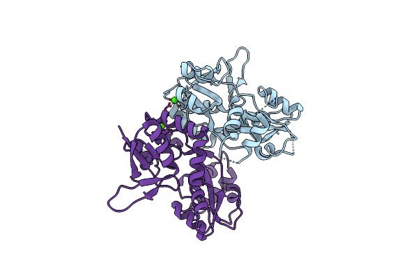 |
Structure Of The Ligand-Binding Core Of The Ionotropic Glutamate Receptor-Like Glurdelta2 In The Apo Form
Organism: Rattus norvegicus
Method: X-RAY DIFFRACTION Resolution:2.75 Å Release Date: 2007-08-07 Classification: RECEPTOR Ligands: CA |
 |
Structure Of The Ligand-Binding Core Of The Ionotropic Glutamate Receptor-Like Glurdelta2 In Complex With D-Serine
Organism: Rattus norvegicus
Method: X-RAY DIFFRACTION Resolution:1.74 Å Release Date: 2007-08-07 Classification: RECEPTOR Ligands: DSN, NA, CL, SCN |
 |
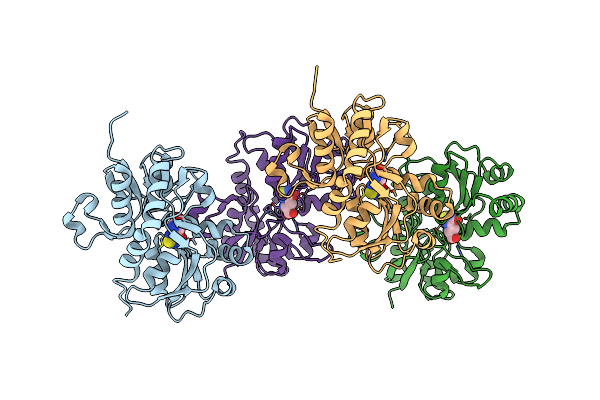
Crystal Structure Of The Glur2 Ligand Binding Core (S1S2J-Y450W) Mutant In Complex With The Partial Agonist Kainic Acid At 2.1 A Resolution
Organism: Rattus norvegicus
Method: X-RAY DIFFRACTION Resolution:2.10 Å Release Date: 2005-08-30 Classification: MEMBRANE PROTEIN Ligands: KAI |
 |
X-Ray Structure Of The Glur2 Ligand-Binding Core (S1S2J) In Complex With (S)-Cpw399 At 1.85 A Resolution.
Organism: Rattus norvegicus
Method: X-RAY DIFFRACTION Resolution:1.80 Å Release Date: 2005-03-22 Classification: MEMBRANE PROTEIN Ligands: CPW |
 |
X-Ray Structure Of The Y702F Mutant Of The Glur2 Ligand-Binding Core (S1S2J) In Complex With (S)-Cpw399 At 2.1 A Resolution.
Organism: Rattus norvegicus
Method: X-RAY DIFFRACTION Resolution:2.10 Å Release Date: 2005-03-22 Classification: MEMBRANE PROTEIN Ligands: CPW |
 |
X-Ray Structure Of The Y702F Mutant Of The Glur2 Ligand-Binding Core (S1S2J) In Complex With Kainate At 1.85 A Resolution
Organism: Rattus norvegicus
Method: X-RAY DIFFRACTION Resolution:1.85 Å Release Date: 2005-03-22 Classification: MEMBRANE PROTEIN Ligands: SO4, KAI |
 |
Crystal Structure Of The Kainate Receptor Glur5 Ligand-Binding Core In Complex With (S)-Glutamate
Organism: Rattus norvegicus
Method: X-RAY DIFFRACTION Resolution:1.95 Å Release Date: 2005-02-01 Classification: MEMBRANE PROTEIN Ligands: SO4, GLU |
 |
Decoding Center & Peptidyl Transferase Center From The X-Ray Structure Of The Thermus Thermophilus 70S Ribosome, Aligned To The Low Resolution Cryo-Em Map Of E.Coli 70S Ribosome
Organism: Escherichia coli
Method: ELECTRON MICROSCOPY Release Date: 2003-04-01 Classification: RIBOSOME |
 |
Docking Of The Modified Rf2 X-Ray Structure Into The Low Resolution Cryo-Em Map Of Rf2 E.Coli 70S Ribosome
Organism: Escherichia coli
Method: ELECTRON MICROSCOPY Resolution:12.80 Å Release Date: 2003-01-14 Classification: TRANSLATION, RIBOSOME |
 |
Structure Of The E. Coli Ribosomal Termination Complex With Release Factor 2
Organism: Escherichia coli
Method: ELECTRON MICROSCOPY Release Date: 2003-01-14 Classification: RIBOSOME |
 |
Organism: Escherichia coli
Method: X-RAY DIFFRACTION Resolution:1.81 Å Release Date: 2002-04-04 Classification: TRANSLATION |