Search Count: 27
 |
Organism: Streptococcus pneumoniae
Method: X-RAY DIFFRACTION Resolution:2.04 Å Release Date: 2017-03-15 Classification: CELL ADHESION Ligands: SO4, NI, CA |
 |
Streptococcus Pneumoniae Glyceraldehyde-3-Phosphate Dehydrogenase (Spgapdh) Crystal Structure
Organism: Streptococcus pneumoniae
Method: X-RAY DIFFRACTION Resolution:2.00 Å Release Date: 2017-01-11 Classification: OXIDOREDUCTASE Ligands: CL, CA, ACY, GOL |
 |
Crystal Structure Of The Autolysin Lyta From Streptococcus Pneumoniae Tigr4
Organism: Streptococcus pneumoniae tigr4
Method: X-RAY DIFFRACTION Resolution:2.10 Å Release Date: 2015-05-27 Classification: HYDROLASE Ligands: GOL, ZN, CHT |
 |
Organism: Streptococcus pneumoniae
Method: SOLUTION NMR Release Date: 2013-11-20 Classification: ZINC-BINDING PROTEIN Ligands: ZN |
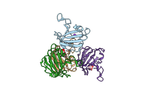 |
Lytm Domain Of Envc, An Activator Of Cell Wall Amidases In Escherichia Coli
Organism: Escherichia coli
Method: X-RAY DIFFRACTION Resolution:1.57 Å Release Date: 2013-07-03 Classification: CELL CYCLE Ligands: IOD, CL, GOL, K |
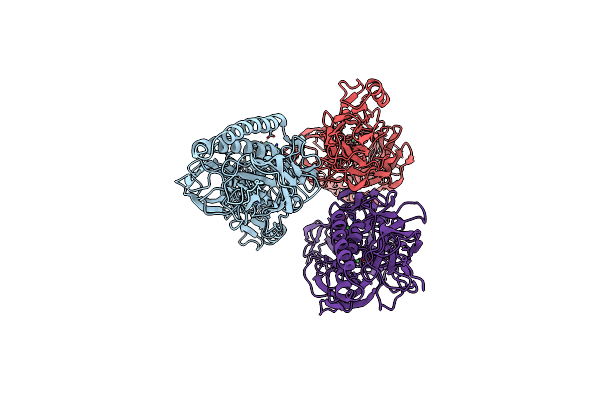 |
Organism: Streptococcus pneumoniae
Method: X-RAY DIFFRACTION Resolution:2.39 Å Release Date: 2011-11-09 Classification: STRUCTURAL PROTEIN Ligands: NI |
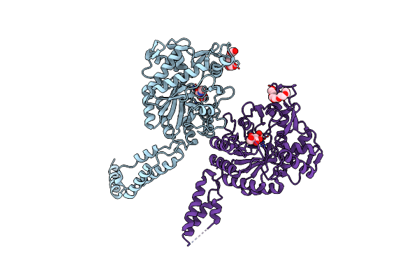 |
Crystal Structure Of The First Gh20 Domain Of A Novel Beta-N-Acetyl-Hexosaminidase Strh From Streptococcus Pneumoniae R6
Organism: Streptococcus pneumoniae
Method: X-RAY DIFFRACTION Resolution:2.10 Å Release Date: 2011-10-26 Classification: HYDROLASE Ligands: NAG, 1PE |
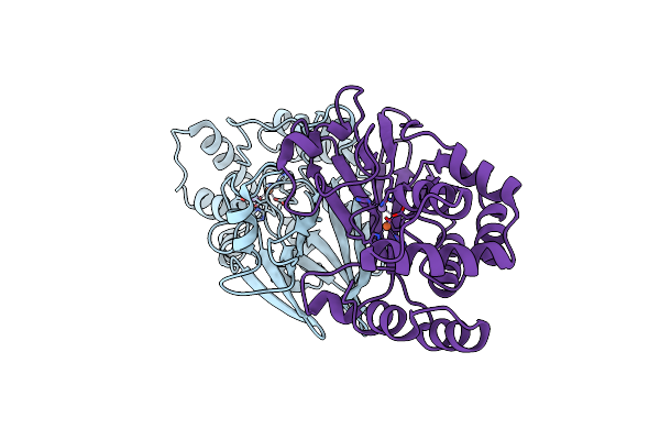 |
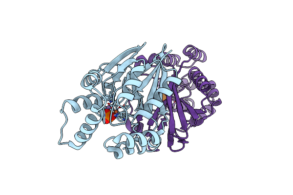
Crystal Structure Of Streptococcal Asymmetric Ap4A Hydrolase And Phosphodiesterase Spr1479/Saph
Organism: Streptococcus pneumoniae
Method: X-RAY DIFFRACTION Resolution:1.90 Å Release Date: 2011-08-24 Classification: HYDROLASE Ligands: MN, FE |
 |
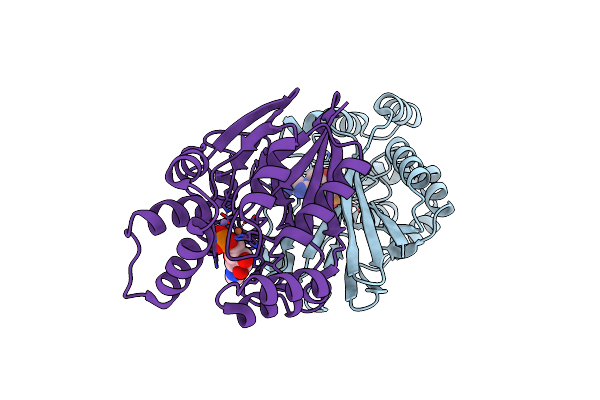
Crystal Structure Of Streptococcal Asymmetric Ap4A Hydrolase And Phosphodiesterase Spr1479/Saph In Complex With Inorganic Phosphate
Organism: Streptococcus pneumoniae
Method: X-RAY DIFFRACTION Resolution:2.31 Å Release Date: 2011-08-24 Classification: HYDROLASE Ligands: PO4, MN, FE |
 |
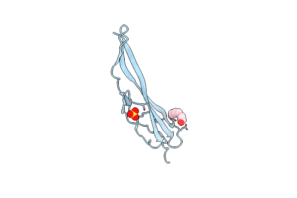
Crystal Structure Of Streptococcal Asymmetric Ap4A Hydrolase And Phosphodiesterase Spr1479/Saph Im Complex With Amp
Organism: Streptococcus pneumoniae
Method: X-RAY DIFFRACTION Resolution:2.20 Å Release Date: 2011-08-24 Classification: HYDROLASE Ligands: AMP, FE, MN |
 |
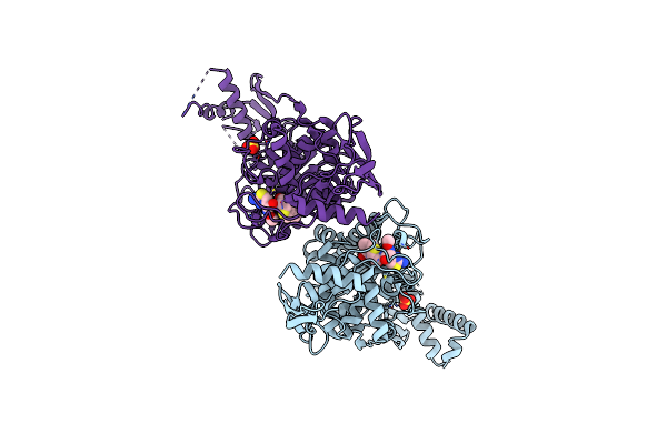
Crystal Structure Of The Mucin-Binding Domain Of Spr1345 From Streptococcus Pneumoniae
Organism: Streptococcus pneumoniae
Method: X-RAY DIFFRACTION Resolution:2.00 Å Release Date: 2011-04-20 Classification: CELL ADHESION Ligands: PGE, SO4 |
 |
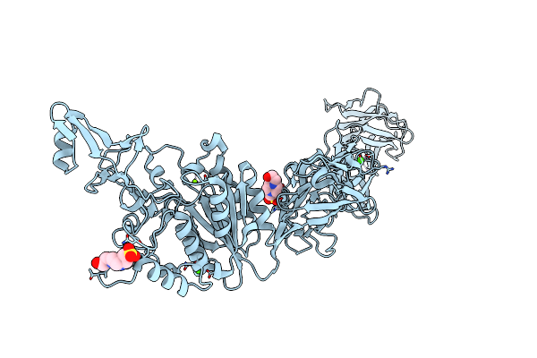
Active Site Restructuring Regulates Ligand Recognition In Class A Penicillin-Binding Proteins
Organism: Streptococcus pneumoniae
Method: X-RAY DIFFRACTION Resolution:3.00 Å Release Date: 2010-05-26 Classification: TRANSFERASE Ligands: CEF, SO4 |
 |
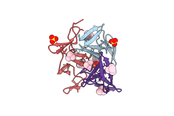
Organism: Streptococcus pneumoniae
Method: X-RAY DIFFRACTION Resolution:1.90 Å Release Date: 2010-01-19 Classification: CELL ADHESION Ligands: MG, CA, EPE |
 |
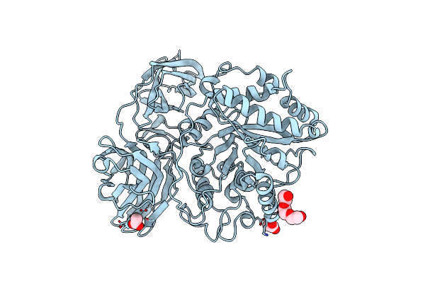
Crystal Structure Of The Choline Binding Domain Of Spr1274 In Streptococcus Pneumoniae
Organism: Streptococcus pneumoniae
Method: X-RAY DIFFRACTION Resolution:2.38 Å Release Date: 2009-08-04 Classification: CHOLINE-BINDING PROTEIN Ligands: PC, SO4 |
 |
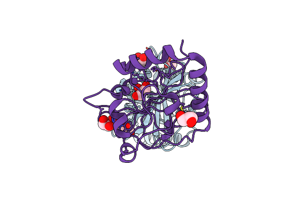
Crystal Structure Of Spr0440 Glycoside Hydrolase Domain, Endo-D From Streptococcus Pneumoniae R6
Organism: Streptococcus pneumoniae
Method: X-RAY DIFFRACTION Resolution:1.87 Å Release Date: 2009-03-17 Classification: HYDROLASE Ligands: ACY, PGE |
 |
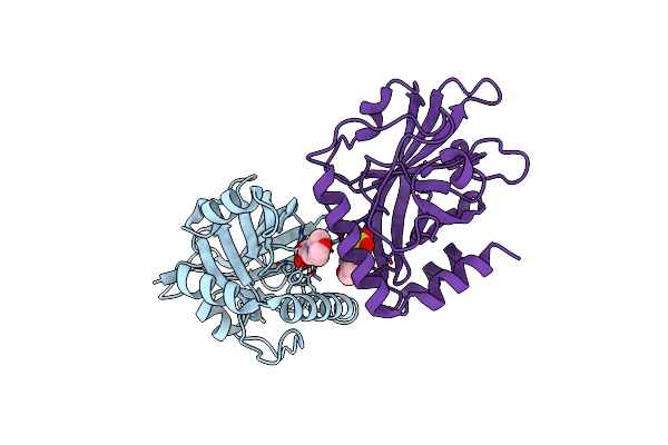
Organism: Streptococcus pneumoniae
Method: X-RAY DIFFRACTION Resolution:1.24 Å Release Date: 2008-12-23 Classification: TRANSFERASE Ligands: GOL |
 |
Organism: Streptococcus pneumoniae
Method: X-RAY DIFFRACTION Resolution:2.14 Å Release Date: 2008-12-23 Classification: TRANSFERASE Ligands: MES |
 |
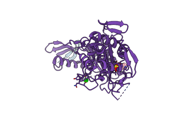
Organism: Streptococcus pneumoniae
Method: X-RAY DIFFRACTION Resolution:2.40 Å Release Date: 2008-08-05 Classification: METAL BINDING PROTEIN |
 |
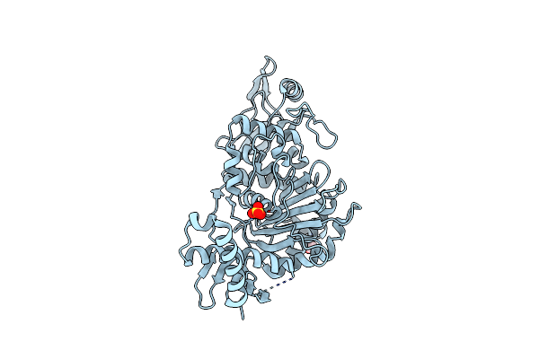
Crystal Structure Of Pbp1A From Drug-Resistant Strain 5204 From Streptococcus Pneumoniae
Organism: Streptococcus pneumoniae
Method: X-RAY DIFFRACTION Resolution:1.90 Å Release Date: 2007-12-25 Classification: TRANSFERASE Ligands: BA, MES |
 |
Active Site Restructuring Regulates Ligand Recognition In Class A Penicillin-Binding Proteins
Organism: Streptococcus pneumoniae
Method: X-RAY DIFFRACTION Resolution:2.39 Å Release Date: 2007-04-03 Classification: TRANSFERASE Ligands: SO4, CL, EDO |

