
Search Count: 25
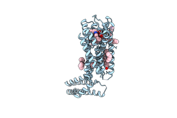 |
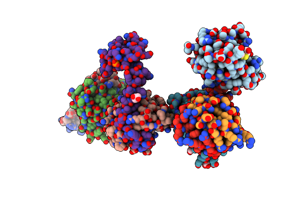
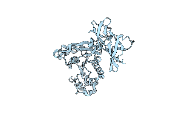
Crystal Structure Of A Peptidergic Gpcr In Complex With A Small Synthetic G Protein-Biased Agonist
Organism: Homo sapiens, Escherichia coli o139:h28 str. e24377a
Method: X-RAY DIFFRACTION Resolution:2.58 Å Release Date: 2024-12-04 Classification: MEMBRANE PROTEIN Ligands: A1D5N, OLA, 1PE |
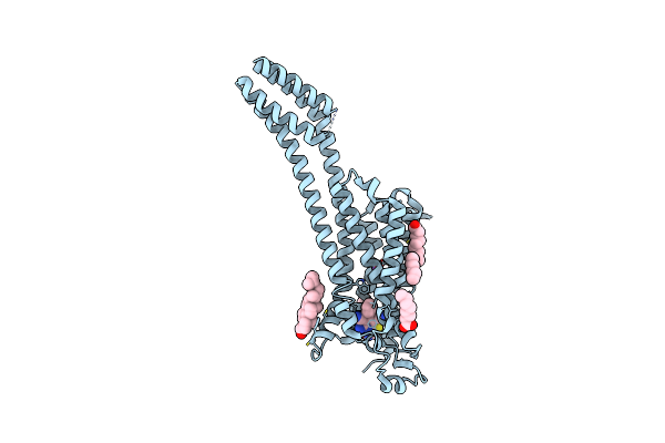 |
Crystal Structure Of The Non-Ribose Partial Agonist Luf5833 Bound To The Adenosine A2A Receptor
Organism: Homo sapiens, Escherichia coli
Method: X-RAY DIFFRACTION Resolution:3.12 Å Release Date: 2021-04-07 Classification: MEMBRANE PROTEIN Ligands: RVZ, OLA, CLR, NA |
 |
Crystal Structure Of Stabilized A2A Adenosine Receptor A2Ar-Star2-Bril In Complex With Chromone 4D
Organism: Homo sapiens, Escherichia coli
Method: X-RAY DIFFRACTION Resolution:1.92 Å Release Date: 2020-09-16 Classification: PROTEIN BINDING Ligands: QGE, NA, CLR, OLA, OLC |
 |
Crystal Structure Of Stabilized A2A Adenosine Receptor A2Ar-Star2-Bril In Complex With Chromone 5D
Organism: Homo sapiens, Escherichia coli
Method: X-RAY DIFFRACTION Resolution:2.13 Å Release Date: 2020-09-16 Classification: PROTEIN BINDING Ligands: QGW, NA, CLR, OLB, OLA, OLC |
 |
Organism: Pyrococcus horikoshii (strain atcc 700860 / dsm 12428 / jcm 9974 / nbrc 100139 / ot-3)
Method: X-RAY DIFFRACTION Resolution:3.50 Å Release Date: 2016-04-20 Classification: TRANSPORT PROTEIN Ligands: ASP, NA |
 |
Crystal Structure Of The Bovine Fructose Transporter Glut5 In An Open Inward-Facing Conformation
Organism: Bos taurus
Method: X-RAY DIFFRACTION Resolution:3.20 Å Release Date: 2015-10-14 Classification: TRANSPORT PROTEIN |
 |
Organism: Rattus norvegicus, Mus musculus
Method: X-RAY DIFFRACTION Resolution:3.27 Å Release Date: 2015-10-07 Classification: TRANSPORT PROTEIN/IMMUNE SYSTEM |
 |
Organism: Pyrococcus horikoshii
Method: X-RAY DIFFRACTION Resolution:4.00 Å Release Date: 2014-09-17 Classification: TRANSPORT PROTEIN |
 |
Organism: Pyrococcus horikoshii
Method: X-RAY DIFFRACTION Resolution:3.41 Å Release Date: 2014-08-13 Classification: TRANSPORT PROTEIN |
 |
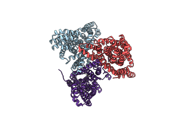
Closed, Apo Inward-Facing State Of The Glutamate Transporter Homologue Gltph
Organism: Pyrococcus horikoshii
Method: X-RAY DIFFRACTION Resolution:3.25 Å Release Date: 2014-06-04 Classification: TRANSPORT PROTEIN Ligands: HG |
 |
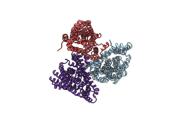
Thallium-Bound Inward-Facing State Of The Glutamate Transporter Homologue Gltph
Organism: Pyrococcus horikoshii
Method: X-RAY DIFFRACTION Resolution:3.75 Å Release Date: 2014-06-04 Classification: TRANSPORT PROTEIN Ligands: TL, HG |
 |
Apo Inward-Facing State Of The Glutamate Transporter Homologue Gltph In Alkali-Free Conditions
Organism: Pyrococcus horikoshii
Method: X-RAY DIFFRACTION Resolution:3.50 Å Release Date: 2014-06-04 Classification: TRANSPORT PROTEIN Ligands: HG |
 |
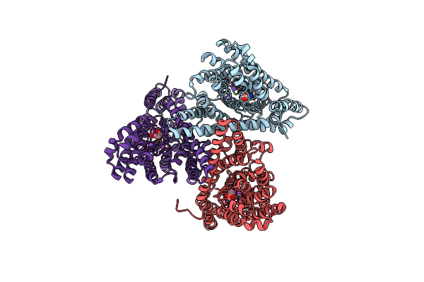
Tl+-Bound Inward-Facing State (Bound Conformation) Of The Glutamate Transporter Homologue Gltph
Organism: Pyrococcus horikoshii
Method: X-RAY DIFFRACTION Resolution:4.08 Å Release Date: 2014-06-04 Classification: TRANSPORT PROTEIN Ligands: HG, TL |
 |
Organism: Pyrococcus horikoshii
Method: X-RAY DIFFRACTION Resolution:3.80 Å Release Date: 2012-02-15 Classification: TRANSPORT PROTEIN Ligands: ASP, NA, HG |
 |
Crystal Structure Of An Asymmetric Trimer Of A Glutamate Transporter Homologue (Gltph)
Organism: Pyrococcus horikoshii
Method: X-RAY DIFFRACTION Resolution:4.66 Å Release Date: 2012-02-15 Classification: TRANSPORT PROTEIN Ligands: ASP, NA |
 |
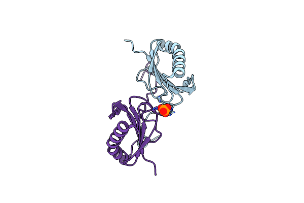
Crystal Structure Of The P1 Bacteriophage Doc Toxin (F68S) In Complex With The Phd Antitoxin (L17M/V39A). Northeast Structural Genomics Targets Er385-Er386
Organism: Bacteriophage p1
Method: X-RAY DIFFRACTION Resolution:2.71 Å Release Date: 2010-08-18 Classification: TOXIN Ligands: CL, HED, GOL, PO4 |
 |
Crystal Structure Of Q83Jn9 From Shigella Flexneri At High Resolution. Northeast Structural Genomics Consortium Target Sfr137.
Organism: Shigella flexneri
Method: X-RAY DIFFRACTION Resolution:1.50 Å Release Date: 2006-10-24 Classification: STRUCTURAL GENOMICS, UNKNOWN FUNCTION Ligands: PO4 |
 |
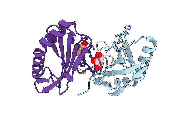
Crystal Structure Of The Bacterial Antitoxin Higa From Escherichia Coli At Ph 8.5. Northeast Structural Genomics Target Er390.
Organism: Escherichia coli
Method: X-RAY DIFFRACTION Resolution:1.63 Å Release Date: 2006-09-26 Classification: DNA BINDING PROTEIN |
 |
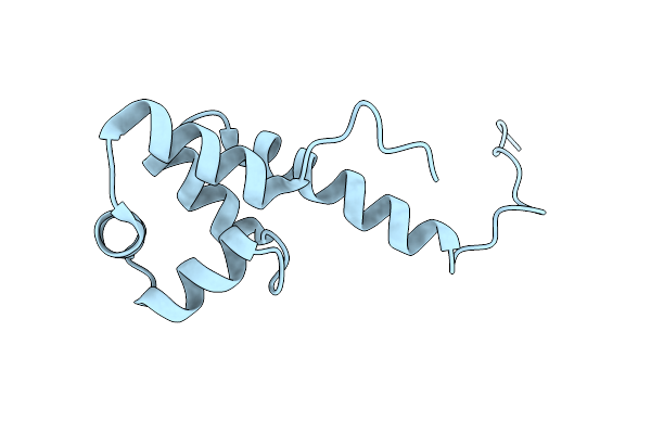
Crystal Structure Of Yeeu From E. Coli. Northeast Structural Genomics Target Er304
Organism: Escherichia coli
Method: X-RAY DIFFRACTION Resolution:2.10 Å Release Date: 2006-07-18 Classification: STRUCTURAL GENOMICS, UNKNOWN FUNCTION Ligands: CL, MG, GOL |
 |
Crystal Structure Of Glcv, The Abc-Atpase Of The Glucose Abc Transporter From Sulfolobus Solfataricus
Organism: Sulfolobus solfataricus
Method: X-RAY DIFFRACTION Resolution:1.45 Å Release Date: 2003-09-30 Classification: TRANSPORT PROTEIN Ligands: IOD |