
Search Count: 13
 |
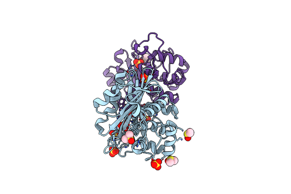
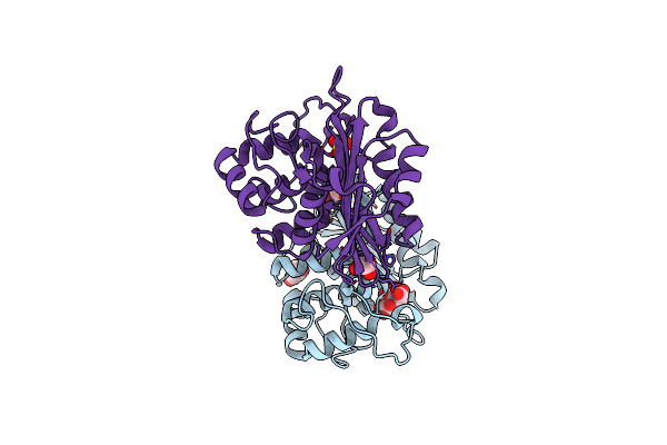
Novel Imidazo[1,2-A]Pyridine Derivatives With Potent Autotaxin/Enpp2 Inhibitor Activity
Organism: Homo sapiens
Method: X-RAY DIFFRACTION Resolution:2.70 Å Release Date: 2017-08-30 Classification: HYDROLASE Ligands: 7HR, IOD, CL, SCN, NI, PO4, K, ZN |
 |
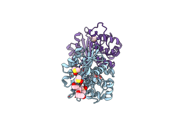
Organism: Pseudomonas aeruginosa
Method: X-RAY DIFFRACTION Resolution:1.90 Å Release Date: 2010-12-08 Classification: HYDROLASE Ligands: SO4, DMS, PG4 |
 |
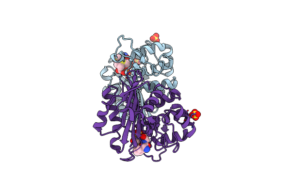
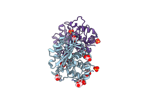
Crystal Structure Of The Class D Beta-Lactamase Oxa-10 At 1.35 A Resolution
Organism: Pseudomonas aeruginosa
Method: X-RAY DIFFRACTION Resolution:1.35 Å Release Date: 2010-12-08 Classification: HYDROLASE Ligands: SO4, PG4, DMS, GOL |
 |
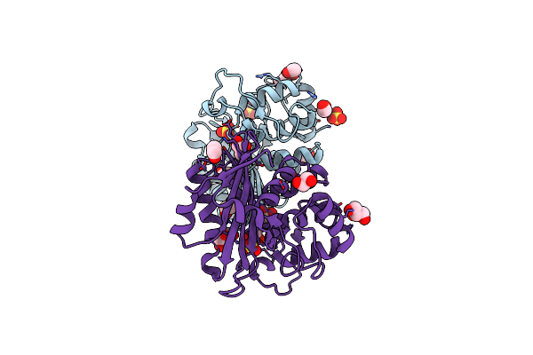
Organism: Pseudomonas aeruginosa
Method: X-RAY DIFFRACTION Resolution:1.79 Å Release Date: 2010-08-25 Classification: HYDROLASE Ligands: ZZ7, SO4 |
 |
Organism: Pseudomonas aeruginosa
Method: X-RAY DIFFRACTION Resolution:2.10 Å Release Date: 2010-08-25 Classification: HYDROLASE Ligands: SO4, GOL, EDO |
 |
Crystal Structure Of The Oxa-10 V117T Mutant At Ph 6.5 Inhibited By A Chloride Ion
Organism: Pseudomonas aeruginosa
Method: X-RAY DIFFRACTION Resolution:1.80 Å Release Date: 2010-05-19 Classification: HYDROLASE Ligands: GOL, CL, CIT, SO4 |
 |
Organism: Pseudomonas aeruginosa
Method: X-RAY DIFFRACTION Resolution:1.80 Å Release Date: 2010-05-19 Classification: HYDROLASE Ligands: SO4, GOL |
 |
Organism: Pseudomonas aeruginosa
Method: X-RAY DIFFRACTION Resolution:2.85 Å Release Date: 2009-11-10 Classification: HYDROLASE/ANTIBIOTIC Ligands: PNM, GOL |
 |
Organism: Pseudomonas aeruginosa
Method: X-RAY DIFFRACTION Resolution:1.90 Å Release Date: 2008-10-28 Classification: HYDROLASE Ligands: SO4, GOL, EDO |
 |
Organism: Pseudomonas aeruginosa
Method: X-RAY DIFFRACTION Resolution:2.70 Å Release Date: 2007-07-03 Classification: HYDROLASE Ligands: SO4, CO |
 |
Organism: Pseudomonas aeruginosa
Method: X-RAY DIFFRACTION Resolution:2.20 Å Release Date: 2007-07-03 Classification: HYDROLASE Ligands: SO4 |
 |
Organism: Pseudomonas aeruginosa
Method: X-RAY DIFFRACTION Resolution:2.50 Å Release Date: 2007-07-03 Classification: HYDROLASE Ligands: SO4 |
 |
Organism: Pseudomonas aeruginosa
Method: X-RAY DIFFRACTION Resolution:2.05 Å Release Date: 2007-07-03 Classification: HYDROLASE Ligands: SO4 |