
Search Count: 36
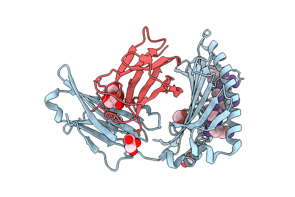 |
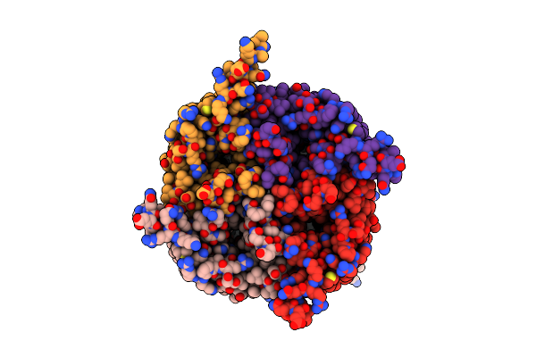
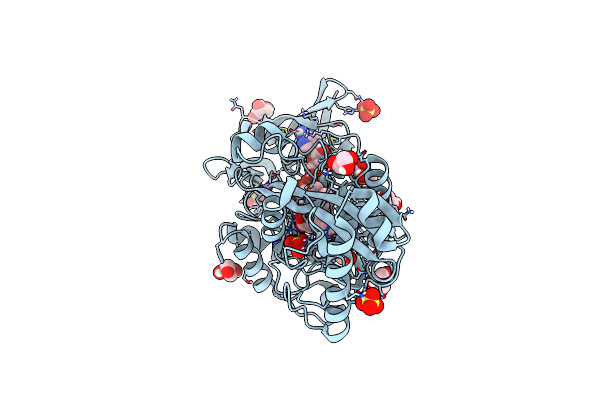
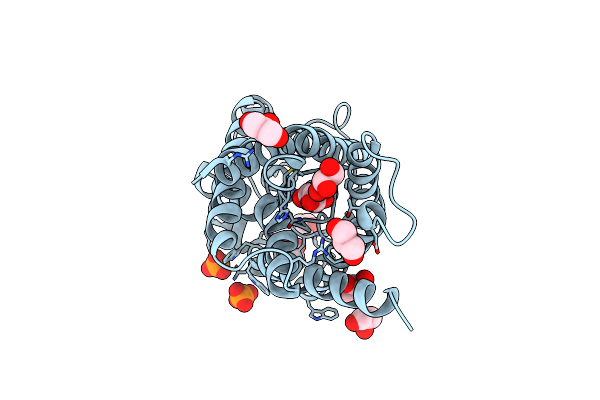
Crystal Structure Of The Great Reed Warbler Mhc Class I In Complex With An 8-Mer Peptide
Organism: Acrocephalus arundinaceus, Legionella
Method: X-RAY DIFFRACTION Release Date: 2025-07-23 Classification: IMMUNE SYSTEM Ligands: GOL |
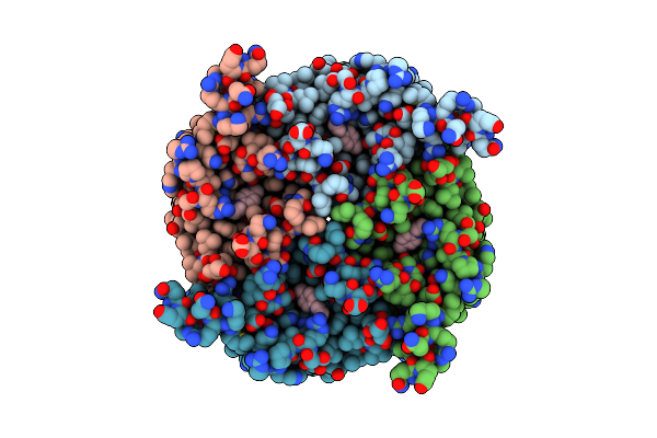 |
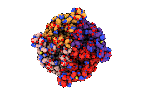
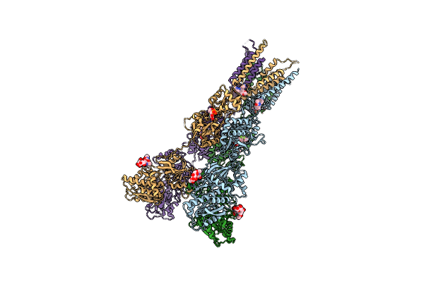
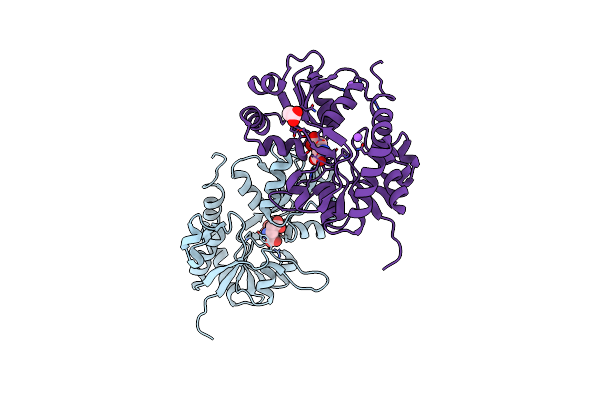
Organism: Homo sapiens
Method: ELECTRON MICROSCOPY Release Date: 2024-01-31 Classification: MEMBRANE PROTEIN Ligands: T60 |
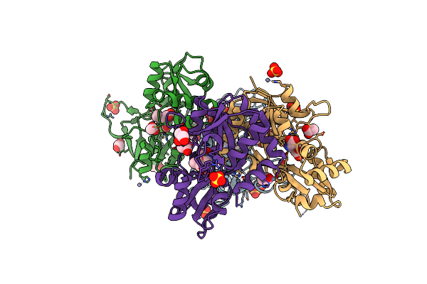 |
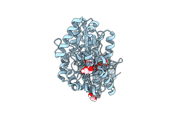
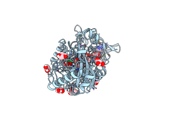
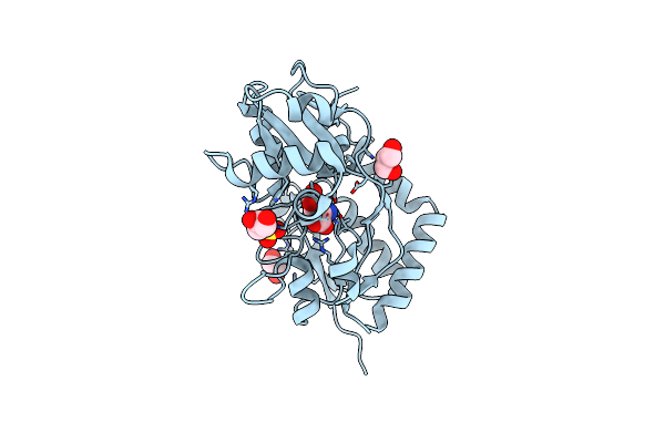
Crystal Structure Of The Kainate Receptor Gluk3-H523A Ligand Binding Domain In Complex With Kainate At 2.7A Resolution
Organism: Rattus norvegicus
Method: X-RAY DIFFRACTION Resolution:2.70 Å Release Date: 2023-12-13 Classification: MEMBRANE PROTEIN Ligands: ACT, ZN, CL, SO4, PEG, KAI, GOL |
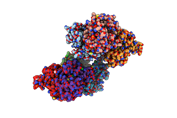 |
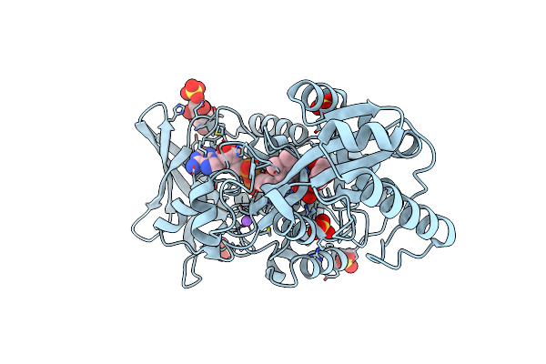
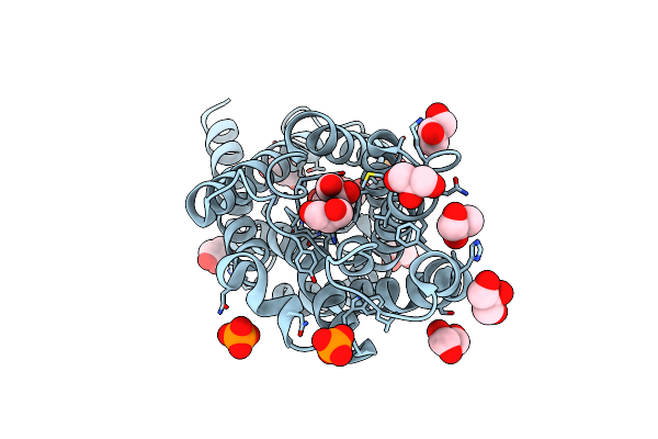
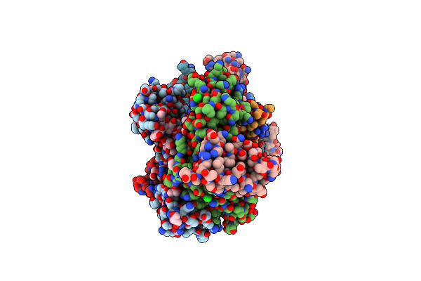
Crystal Structure Of The Kainate Receptor Gluk3-H523A Ligand Binding Domain In Complex With Kainate And The Positive Allosteric Modulator Bpam344 At 2.9A Resolution
Organism: Rattus norvegicus
Method: X-RAY DIFFRACTION Resolution:2.90 Å Release Date: 2023-12-13 Classification: MEMBRANE PROTEIN Ligands: 2J9, ZN, GOL, CL, SO4, KAI, ACT |
 |
Organism: Homo sapiens
Method: ELECTRON MICROSCOPY Release Date: 2023-02-15 Classification: MEMBRANE PROTEIN Ligands: GOL |
 |
Organism: Homo sapiens
Method: ELECTRON MICROSCOPY Release Date: 2023-02-15 Classification: MEMBRANE PROTEIN |
 |
Organism: Shewanella oneidensis (strain mr-1)
Method: X-RAY DIFFRACTION Resolution:1.10 Å Release Date: 2021-03-03 Classification: OXIDOREDUCTASE Ligands: ADP, CL, GOL |
 |
Organism: Shewanella oneidensis (strain mr-1)
Method: X-RAY DIFFRACTION Resolution:2.56 Å Release Date: 2021-03-03 Classification: OXIDOREDUCTASE Ligands: FAD, GOL, CL, SO4, NA |
 |
Organism: Shewanella oneidensis (strain mr-1)
Method: X-RAY DIFFRACTION Resolution:1.56 Å Release Date: 2021-03-03 Classification: OXIDOREDUCTASE Ligands: FAD, URO, GOL, SO4, CL, NA |
 |
Organism: Shewanella oneidensis (strain mr-1)
Method: X-RAY DIFFRACTION Resolution:1.40 Å Release Date: 2021-03-03 Classification: OXIDOREDUCTASE Ligands: GOL, FAD, MWQ, NA, CL |
 |
Structure Of Glua2Cryst In Complex The Antagonist Zk200775 And The Negative Allosteric Modulator Gyki53655 At 4.65 A Resolution
Organism: Rattus norvegicus
Method: X-RAY DIFFRACTION Resolution:4.65 Å Release Date: 2020-06-24 Classification: MEMBRANE PROTEIN Ligands: GYK, ZK1 |
 |
Organism: Homo sapiens
Method: X-RAY DIFFRACTION Resolution:1.90 Å Release Date: 2019-11-27 Classification: MEMBRANE PROTEIN Ligands: GOL, PO4 |
 |
Organism: Homo sapiens
Method: X-RAY DIFFRACTION Resolution:2.20 Å Release Date: 2019-11-27 Classification: MEMBRANE PROTEIN Ligands: GOL, PO4 |
 |
Structure Of Glua2-N775S Ligand-Binding Domain (S1S2J) In Complex With Glutamate And Rubidium Bromide At 1.75 A Resolution
Organism: Rattus norvegicus
Method: X-RAY DIFFRACTION Resolution:1.75 Å Release Date: 2019-05-15 Classification: MEMBRANE PROTEIN Ligands: GLU, SO4, GOL, BR |
 |
Structure Of Glua2O Ligand-Binding Domain (S1S2J) In Complex With Glutamate And Sodium Bromide At 1.95 A Resolution
Organism: Rattus norvegicus
Method: X-RAY DIFFRACTION Resolution:1.95 Å Release Date: 2019-05-15 Classification: MEMBRANE PROTEIN Ligands: GLU, BR, GOL, NA, ACT |
 |
Organism: Homo sapiens, Staphylococcus aureus
Method: X-RAY DIFFRACTION Resolution:2.50 Å Release Date: 2018-06-20 Classification: IMMUNE SYSTEM Ligands: PEG, GOL, CA, CL |
 |
Crystal Structure Of The Kainate Receptor Gluk3 Ligand Binding Domain In Complex With (S)-1-[2'-Amino-2'-Carboxyethyl]-6-Methyl-5,7-Dihydropyrrolo[3,4-D]Pyrimidin-2,4(1H,3H)-Dione At Resolution 2.4A
Organism: Rattus norvegicus
Method: X-RAY DIFFRACTION Resolution:2.40 Å Release Date: 2018-02-28 Classification: MEMBRANE PROTEIN Ligands: ZN, CG8, CL, SO4 |
 |
Crystal Structure Of The Kainate Receptor Gluk3 Ligand Binding Domain In Complex With (S)-1-[2-Amino-2-Carboxyethyl]-5,7-Dihydrothieno[3,4-D]Pyrimidin-2,4(1H,3H)-Dione At Resolution 2.6A
Organism: Rattus norvegicus
Method: X-RAY DIFFRACTION Resolution:2.60 Å Release Date: 2018-02-28 Classification: MEMBRANE PROTEIN Ligands: CGW, K, CL |
 |
Structure Of Gluk1 Ligand-Binding Domain (S1S2) In Complex With Lm-12B At 2.05 A Resolution
Organism: Rattus norvegicus
Method: X-RAY DIFFRACTION Resolution:2.05 Å Release Date: 2017-07-26 Classification: MEMBRANE PROTEIN Ligands: SO4, CL, GOL, 8VE, ACT |
 |
Structure Of Gluk1 Ligand-Binding Domain (S1S2) In Complex With Cip-As At 2.85 A Resolution
Organism: Rattus norvegicus
Method: X-RAY DIFFRACTION Resolution:2.85 Å Release Date: 2017-07-26 Classification: MEMBRANE PROTEIN Ligands: CL, 8VN, GOL, SO4 |