
Search Count: 10
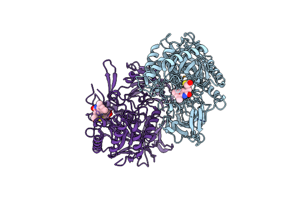 |
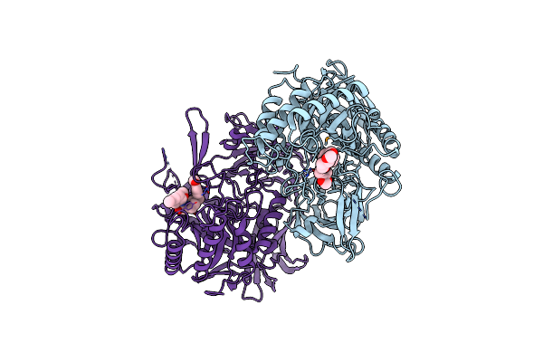
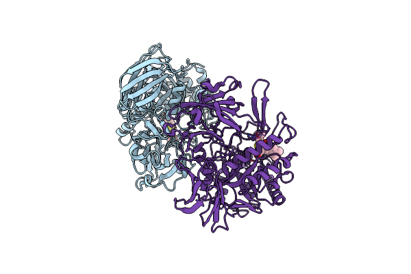
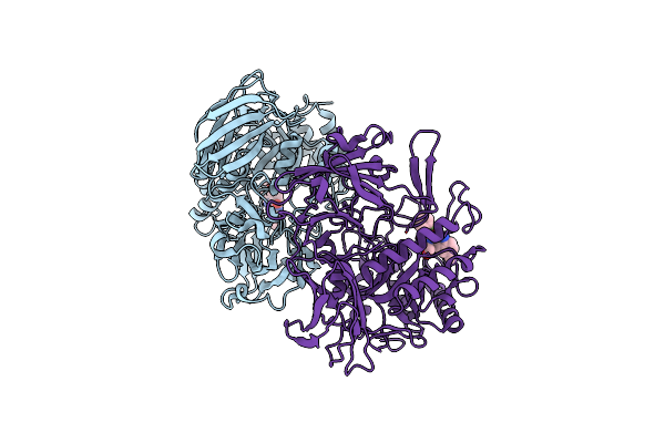
Structure Of E. Coli Beta-Glucuronidase Bound With A Novel, Potent Inhibitor 1-((6,8-Dimethyl-2-Oxo-1,2-Dihydroquinolin-3-Yl)Methyl)-1-(2-Hydroxyethyl)-3-(4-Hydroxyphenyl)Thiourea
Organism: Escherichia coli
Method: X-RAY DIFFRACTION Resolution:2.39 Å Release Date: 2015-10-14 Classification: Hydrolase/Hydrolase Inhibitor Ligands: 57Z |
 |
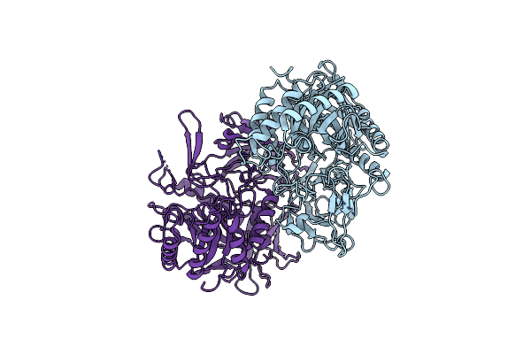
Crystal Structure Of Streptococcus Agalactiae Beta-Glucuronidase In Space Group I222
Organism: Streptococcus agalactiae
Method: X-RAY DIFFRACTION Resolution:2.59 Å Release Date: 2014-09-17 Classification: HYDROLASE Ligands: MG |
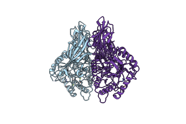 |
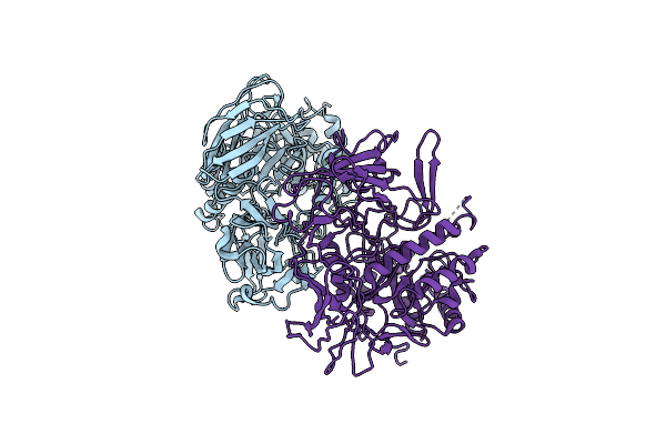
Crystal Structure Of Streptococcus Agalactiae Beta-Glucuronidase In Space Group P21212
Organism: Streptococcus agalactiae
Method: X-RAY DIFFRACTION Resolution:2.29 Å Release Date: 2014-09-17 Classification: HYDROLASE Ligands: MG |
 |
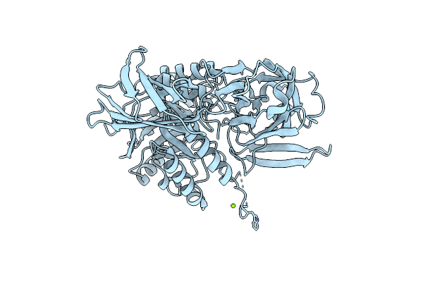
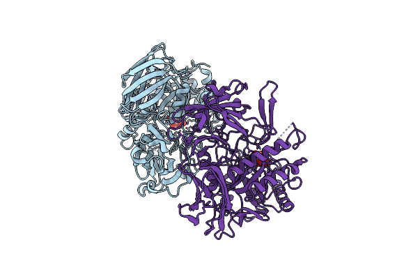
Organism: Clostridium perfringens, Escherichia coli
Method: X-RAY DIFFRACTION Resolution:2.26 Å Release Date: 2014-09-17 Classification: HYDROLASE |
 |
Structure Of E. Coli Beta-Glucuronidase Bound With A Novel, Potent Inhibitor 2-[4-(1,3-Benzodioxol-5-Ylmethyl)Piperazin-1-Yl]-N-[(1S,2S,5S)-2,5-Dimethoxycyclohexyl]Acetamide
Organism: Escherichia coli
Method: X-RAY DIFFRACTION Resolution:2.83 Å Release Date: 2013-08-28 Classification: hydrolase/hydrolase inhibitor Ligands: 1KV |
 |
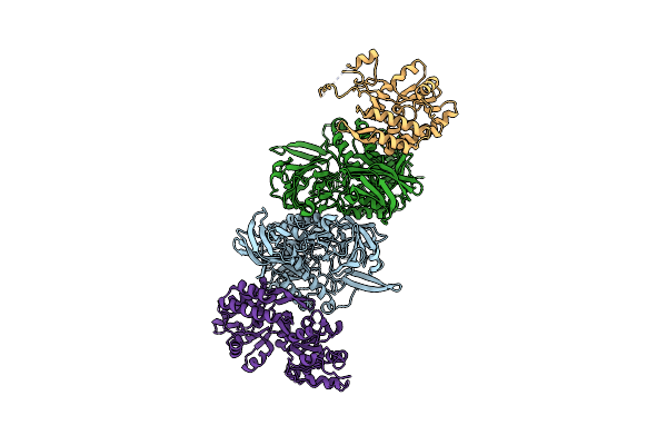
Organism: Escherichia coli
Method: X-RAY DIFFRACTION Resolution:2.50 Å Release Date: 2010-11-17 Classification: HYDROLASE |
 |
Crystal Structure Of Selenomethionine Substituted E. Coli Beta-Glucuronidase
Organism: Escherichia coli
Method: X-RAY DIFFRACTION Resolution:2.90 Å Release Date: 2010-11-17 Classification: HYDROLASE |
 |
Crystal Structure Of E. Coli Beta-Glucuronidase With The Glucaro-D-Lactam Inhibitor Bound
Organism: Escherichia coli
Method: X-RAY DIFFRACTION Resolution:2.39 Å Release Date: 2010-11-17 Classification: HYDROLASE/HYDROLASE INHIBITOR Ligands: EVA |
 |
Structure Of E. Coli Beta-Glucuronidase Bound With A Novel, Potent Inhibitor 1-((6,7-Dimethyl-2-Oxo-1,2-Dihydroquinolin-3-Yl)Methyl)-1-(2-Hydroxyethyl)-3-(3-Methoxyphenyl)Thiourea
Organism: Escherichia coli
Method: X-RAY DIFFRACTION Resolution:2.26 Å Release Date: 2010-11-17 Classification: HYDROLASE/HYDROLASE INHIBITOR Ligands: Z77 |
 |
Structure Of E. Coli Beta-Glucuronidase Bound With A Novel, Potent Inhibitor 3-(2-Fluorophenyl)-1-(2-Hydroxyethyl)-1-((6-Methyl-2-Oxo-1,2-Dihydroquinolin-3-Yl)Methyl)Urea
Organism: Escherichia coli
Method: X-RAY DIFFRACTION Resolution:2.43 Å Release Date: 2010-11-17 Classification: HYDROLASE/HYDROLASE INHIBITOR Ligands: Z78 |