
Search Count: 214
 |
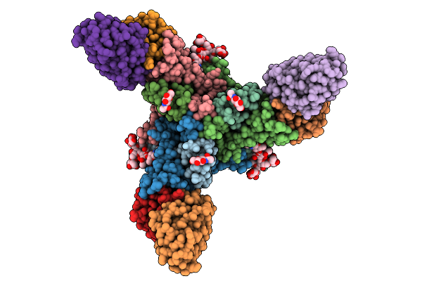
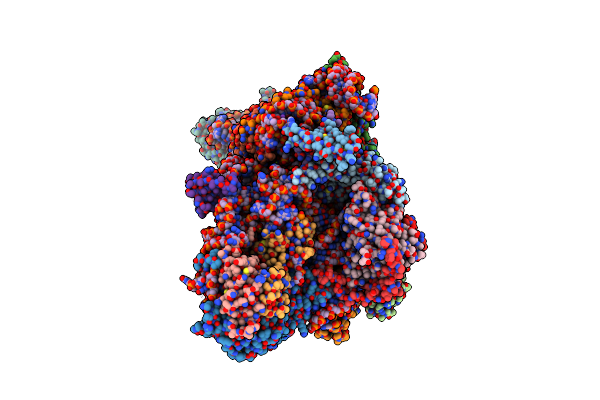
Local Refinement Of The Sars-Cov-2 Ba.2.86 Rbd In Complex With Tri2-2 Minibinder
Organism: Severe acute respiratory syndrome coronavirus 2, Synthetic construct
Method: ELECTRON MICROSCOPY Release Date: 2026-01-21 Classification: VIRAL PROTEIN Ligands: NAG |
 |
Organism: Severe acute respiratory syndrome coronavirus 2
Method: ELECTRON MICROSCOPY Release Date: 2026-01-21 Classification: VIRAL PROTEIN Ligands: NAG |
 |
Organism: Severe acute respiratory syndrome coronavirus 2, Synthetic construct
Method: ELECTRON MICROSCOPY Resolution:2.40 Å Release Date: 2026-01-21 Classification: VIRAL PROTEIN Ligands: NAG |
 |
Organism: Streptomyces coelicolor
Method: ELECTRON MICROSCOPY Release Date: 2025-12-17 Classification: TOXIN Ligands: A2G, A1CAY, A1CAZ |
 |
Organism: Mus musculus, Orthomarburgvirus marburgense
Method: ELECTRON MICROSCOPY Resolution:2.60 Å Release Date: 2025-11-26 Classification: VIRAL PROTEIN Ligands: NAG |
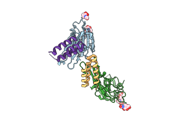 |
Designed Miniproteins Potently Inhibit And Protect Against Mers-Cov. Mers-Cov S In Complex With Miniprotein Cb3_Gggsgggs_Sb175, Linker 7 (Local Refinement Of Two Rbds And 2 Miniproteins)
Organism: Middle east respiratory syndrome-related coronavirus, Synthetic construct
Method: ELECTRON MICROSCOPY Release Date: 2025-10-15 Classification: VIRAL PROTEIN Ligands: NAG |
 |
Structure Of The Rattus Norvegicus Ace2 Receptor Bound Hsitaly2011 Rbd Complex
Organism: Rattus norvegicus, Merbecovirus
Method: ELECTRON MICROSCOPY Release Date: 2025-08-27 Classification: HYDROLASE/VIRAL PROTEIN Ligands: NAG, ZN |
 |
Organism: Eptesicus fuscus, Merbecovirus
Method: ELECTRON MICROSCOPY Release Date: 2025-08-27 Classification: HYDROLASE/VIRAL PROTEIN Ligands: NAG, ZN |
 |
Molecular Basis Of Pathogenicity Of The Recently Emerged Fcov-23 Coronavirus. Complex Of Fapn With Fcov-23 Rbd
Organism: Felis catus, Feline coronavirus
Method: ELECTRON MICROSCOPY Release Date: 2025-07-09 Classification: VIRAL PROTEIN/HYDROLASE Ligands: NAG, ZN |
 |
Molecular Basis Of Pathogenicity Of The Recently Emerged Fcov-23 Coronavirus. Fcov-23 S Short
Organism: Feline coronavirus
Method: ELECTRON MICROSCOPY Release Date: 2025-07-09 Classification: VIRAL PROTEIN Ligands: NAG, PAM |
 |
Molecular Basis Of Pathogenicity Of The Recently Emerged Fcov-23 Coronavirus. Fcov-23 S Do In Proximal Conformation (Local Refinement)
Organism: Feline coronavirus
Method: ELECTRON MICROSCOPY Release Date: 2025-07-09 Classification: VIRAL PROTEIN Ligands: NAG |
 |
Molecular Basis Of Pathogenicity Of The Recently Emerged Fcov-23 Coronavirus. Fcov-23 S Long With Do In Swung-Out Conformation
Organism: Feline coronavirus
Method: ELECTRON MICROSCOPY Release Date: 2025-07-09 Classification: VIRAL PROTEIN Ligands: NAG, PAM |
 |
Molecular Basis Of Pathogenicity Of The Recently Emerged Fcov-23 Coronavirus. Fcov-23 S Long Domain 0 In Swung-Out Conformation (Local Refinement)
Organism: Feline coronavirus
Method: ELECTRON MICROSCOPY Release Date: 2025-07-09 Classification: VIRAL PROTEIN Ligands: NAG |
 |
Molecular Basis Of Pathogenicity Of The Recently Emerged Fcov-23 Coronavirus. Fcov-23 S Long With Do In Mixed Conformations (Global Refinement).
Organism: Feline coronavirus
Method: ELECTRON MICROSCOPY Release Date: 2025-07-09 Classification: VIRAL PROTEIN Ligands: NAG, PAM |
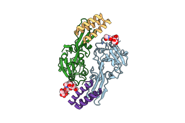 |
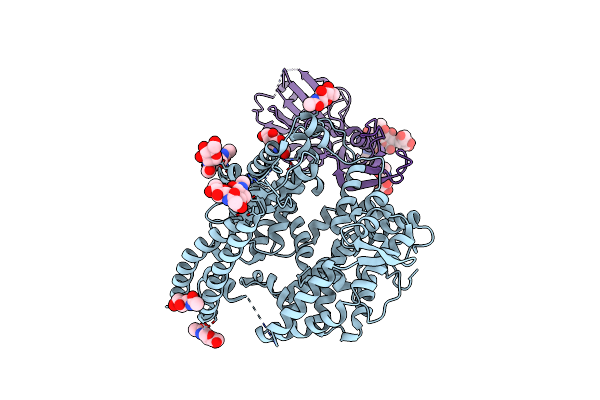
Designed Miniproteins Potently Inhibit And Protect Against Mers-Cov. Crystal Structure Of Mers-Cov S Rbd In Complex With Miniprotein Cb3
Organism: Middle east respiratory syndrome-related coronavirus, Synthetic construct
Method: X-RAY DIFFRACTION Resolution:1.85 Å Release Date: 2025-06-18 Classification: VIRAL PROTEIN Ligands: NAG |
 |
Sars-Cov-2 Nsp1 Bound To The Rhinolophus Lepidus 40S Ribosomal Subunit (Local Refinement Of The 40S Body)
Organism: Severe acute respiratory syndrome coronavirus 2, Homo sapiens, Rhinolophus lepidus
Method: ELECTRON MICROSCOPY Resolution:2.10 Å Release Date: 2025-06-11 Classification: RIBOSOME Ligands: MG, K, ZN |
 |
Sars-Cov-2 Nsp1 Bound To The Rhinolophus Lepidus 40S Ribosome (Local Refinement Of The 40S Head)
Organism: Rhinolophus lepidus
Method: ELECTRON MICROSCOPY Resolution:2.10 Å Release Date: 2025-06-11 Classification: RIBOSOME Ligands: MG, ZN, K |
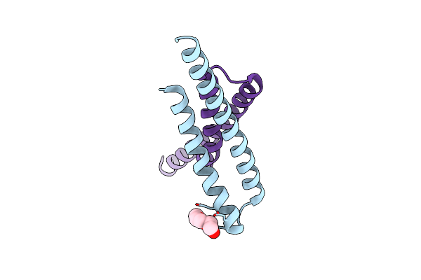 |
Organism: Synthetic construct
Method: X-RAY DIFFRACTION Resolution:2.10 Å Release Date: 2025-05-21 Classification: DE NOVO PROTEIN Ligands: PG4 |
 |
Organism: Merbecovirus, Pteronotus davyi
Method: ELECTRON MICROSCOPY Release Date: 2025-03-05 Classification: VIRAL PROTEIN Ligands: NAG |
 |
Organism: Pipistrellus bat coronavirus hku5
Method: ELECTRON MICROSCOPY Resolution:2.00 Å Release Date: 2025-02-26 Classification: VIRAL PROTEIN Ligands: NAG, ZN, FOL, EIC |