
Search Count: 15
 |
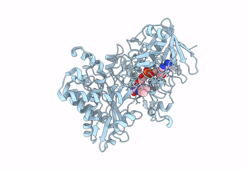
Organism: Acanthamoeba polyphaga mimivirus
Method: ELECTRON MICROSCOPY Release Date: 2022-08-10 Classification: VIRAL PROTEIN Ligands: FAD |
 |
Organism: Acanthamoeba polyphaga mimivirus
Method: ELECTRON MICROSCOPY Release Date: 2022-08-10 Classification: VIRAL PROTEIN Ligands: FAD |
 |
Organism: Acanthamoeba polyphaga mimivirus
Method: ELECTRON MICROSCOPY Release Date: 2022-08-10 Classification: VIRAL PROTEIN Ligands: FAD |
 |
Organism: Acanthamoeba polyphaga mimivirus
Method: ELECTRON MICROSCOPY Release Date: 2022-08-10 Classification: VIRAL PROTEIN Ligands: FAD |
 |
Organism: Escherichia virus m13
Method: ELECTRON MICROSCOPY Release Date: 2021-04-14 Classification: DNA |
 |
Pseudoatomic Model Of The Pre-Fusion Conformation Of Glycoprotein B Of Herpes Simplex Virus 1
Organism: Human herpesvirus 1
Method: ELECTRON MICROSCOPY Release Date: 2020-10-07 Classification: VIRAL PROTEIN |
 |
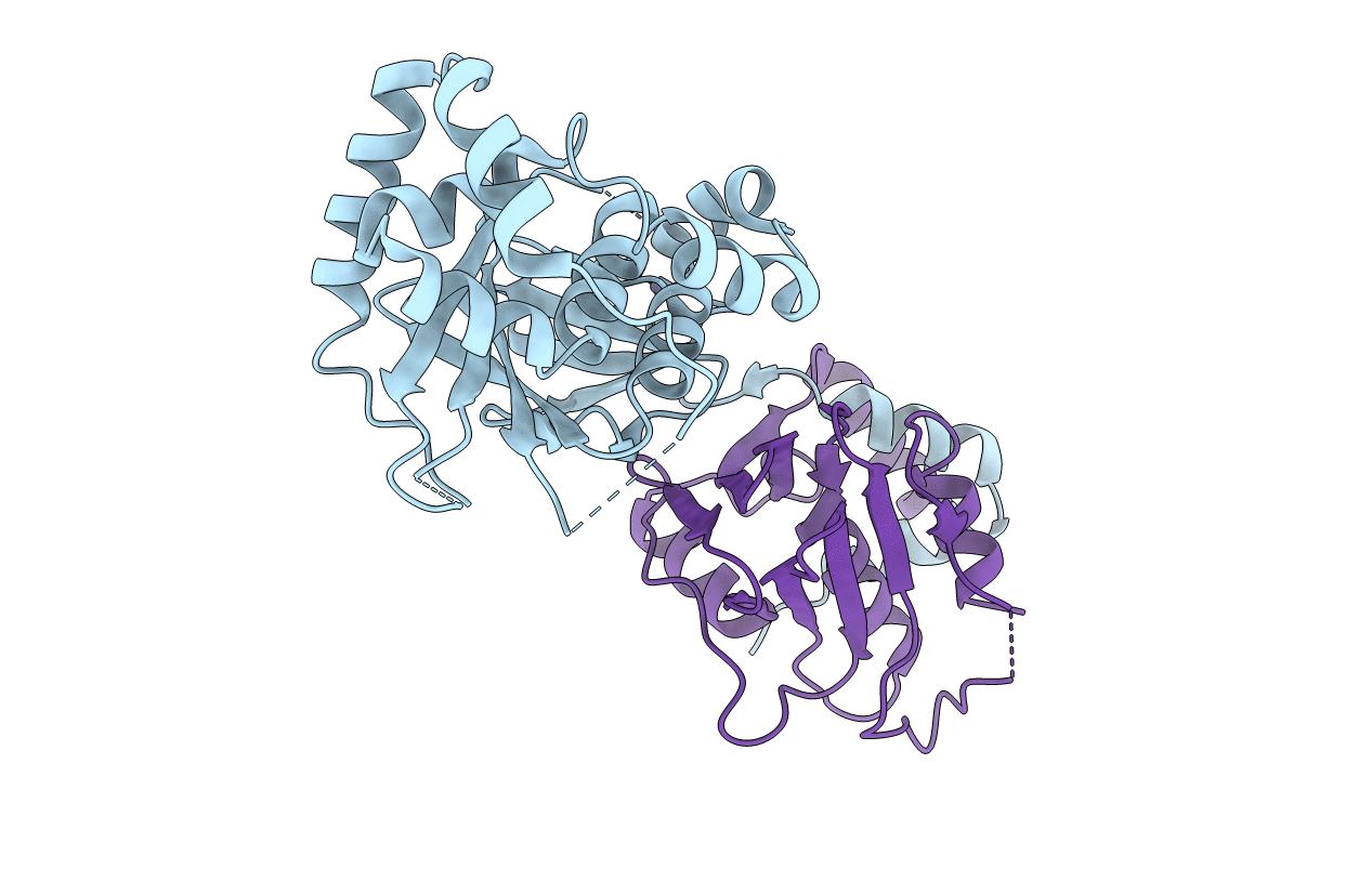
Pseudorabies Virus (Prv) Nuclear Egress Complex Proteins Fitted As A Hexameric Lattice Into A Sub-Tomogram Average Derived From Focused- Ion Beam Milled Lamellae Electron Cryo-Microscopic Data
Organism: Suid herpesvirus 1
Method: ELECTRON MICROSCOPY Resolution:35.00 Å Release Date: 2016-03-16 Classification: VIRAL PROTEIN Ligands: ZN, CL |
 |
Organism: Suid herpesvirus 1
Method: X-RAY DIFFRACTION Resolution:2.90 Å Release Date: 2015-12-23 Classification: VIRAL PROTEIN Ligands: ZN, CL |
 |
Organism: Caenorhabditis elegans
Method: ELECTRON MICROSCOPY Resolution:22.20 Å Release Date: 2014-06-04 Classification: CELL ADHESION |
 |
Organism: Escherichia coli
Method: ELECTRON MICROSCOPY Resolution:8.00 Å Release Date: 2012-12-12 Classification: CHAPERONE Ligands: ATP, PO4, MG |
 |
Organism: Escherichia coli
Method: ELECTRON MICROSCOPY Resolution:8.00 Å Release Date: 2012-12-12 Classification: CHAPERONE Ligands: ATP, PO4, MG |
 |
Organism: Escherichia coli
Method: ELECTRON MICROSCOPY Resolution:8.50 Å Release Date: 2012-12-12 Classification: CHAPERONE Ligands: PO4, MG, ATP |
 |
Organism: Escherichia coli
Method: ELECTRON MICROSCOPY Resolution:8.50 Å Release Date: 2012-12-12 Classification: CHAPERONE Ligands: MG, PO4, ATP |
 |
Organism: Escherichia coli
Method: ELECTRON MICROSCOPY Resolution:8.50 Å Release Date: 2012-12-12 Classification: CHAPERONE Ligands: ATP, MG, PO4 |
 |
Organism: Escherichia coli
Method: ELECTRON MICROSCOPY Resolution:8.50 Å Release Date: 2012-12-12 Classification: CHAPERONE Ligands: PO4, MG, ATP |