
Search Count: 15
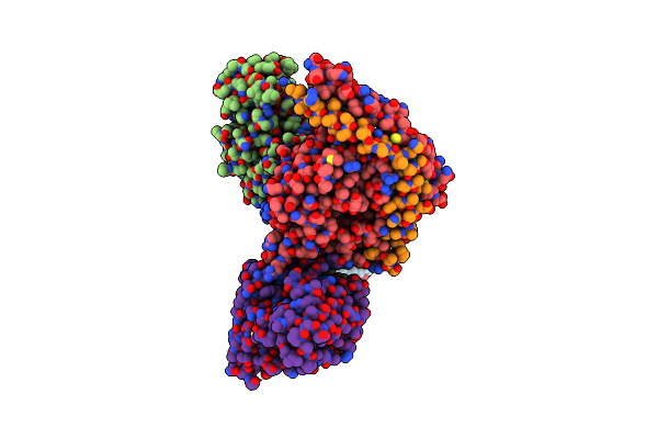 |
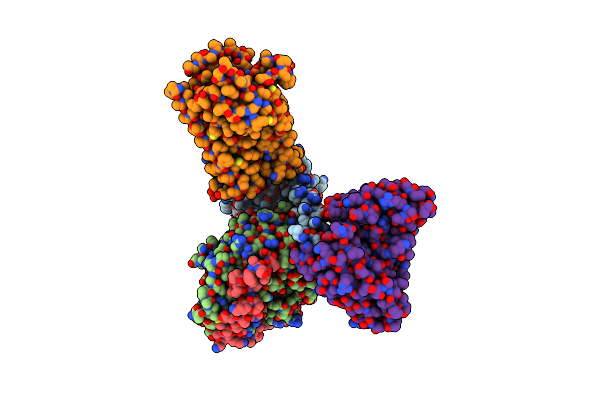
Cryoem Structure Of Delta Opioid Receptor Bound To G Proteins And A Partial Agonist
Organism: Homo sapiens, Mus musculus
Method: ELECTRON MICROSCOPY Resolution:2.80 Å Release Date: 2025-04-02 Classification: MEMBRANE PROTEIN Ligands: A1AWC |
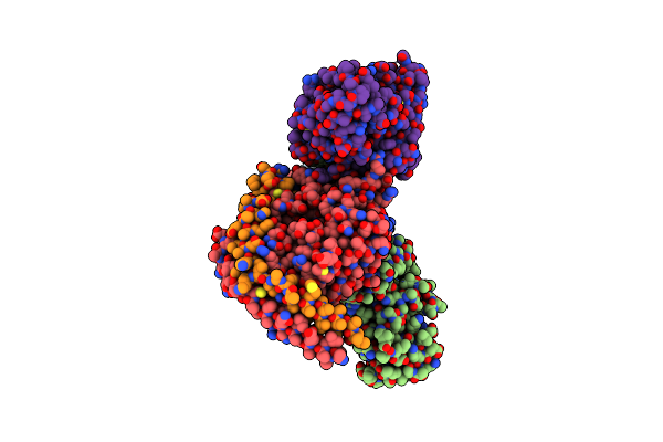 |
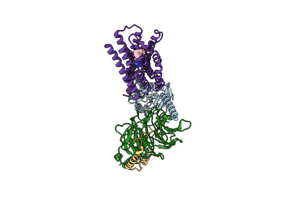
Cryoem Structure Of Delta Opioid Receptor Bound To G Proteins And A Full Bitopic Agonist
Organism: Homo sapiens, Mus musculus
Method: ELECTRON MICROSCOPY Resolution:2.62 Å Release Date: 2025-04-02 Classification: MEMBRANE PROTEIN Ligands: A1AWD |
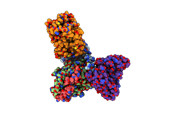 |
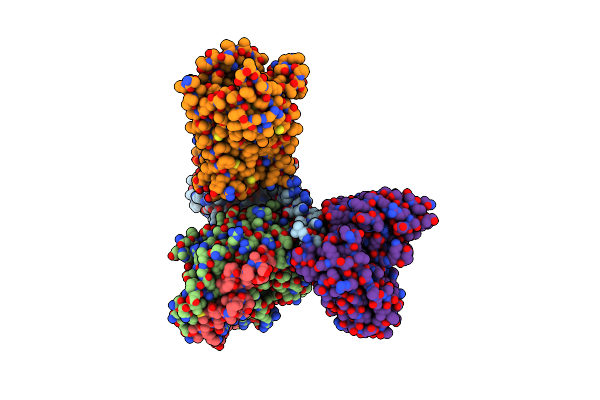
Organism: Homo sapiens, Mus musculus
Method: ELECTRON MICROSCOPY Release Date: 2025-03-05 Classification: SIGNALING PROTEIN Ligands: A1AIW |
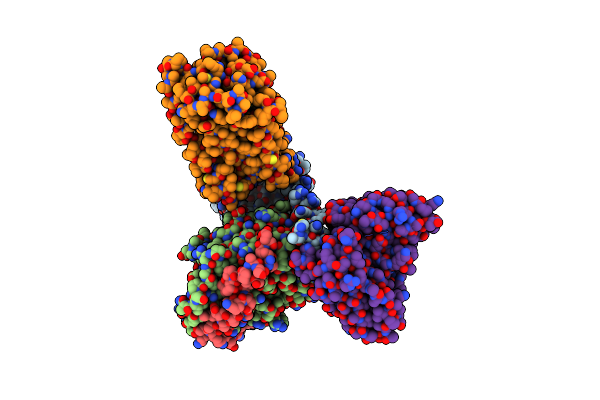 |
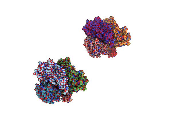
Organism: Homo sapiens, Mus musculus
Method: ELECTRON MICROSCOPY Release Date: 2025-03-05 Classification: SIGNALING PROTEIN Ligands: KCA |
 |
Organism: Homo sapiens, Mus musculus
Method: ELECTRON MICROSCOPY Release Date: 2024-09-11 Classification: SIGNALING PROTEIN/IMMUNE SYSTEM Ligands: A1AQ2 |
 |
Organism: Homo sapiens, Mus musculus, Synthetic construct
Method: ELECTRON MICROSCOPY Release Date: 2023-12-06 Classification: MEMBRANE PROTEIN |
 |
Organism: Homo sapiens, Mus musculus
Method: ELECTRON MICROSCOPY Release Date: 2022-12-07 Classification: MEMBRANE PROTEIN Ligands: KZR |
 |
Organism: Homo sapiens, Mus musculus
Method: ELECTRON MICROSCOPY Release Date: 2022-05-04 Classification: MEMBRANE PROTEIN Ligands: L0X |
 |
Organism: Trametes multicolor
Method: X-RAY DIFFRACTION Resolution:1.70 Å Release Date: 2008-12-02 Classification: OXIDOREDUCTASE Ligands: FAD |
 |
Organism: Trametes multicolor
Method: X-RAY DIFFRACTION Resolution:2.10 Å Release Date: 2008-12-02 Classification: OXIDOREDUCTASE Ligands: FAD |
 |
Organism: Trametes multicolor
Method: X-RAY DIFFRACTION Resolution:1.90 Å Release Date: 2008-12-02 Classification: OXIDOREDUCTASE Ligands: FAD, MES |
 |
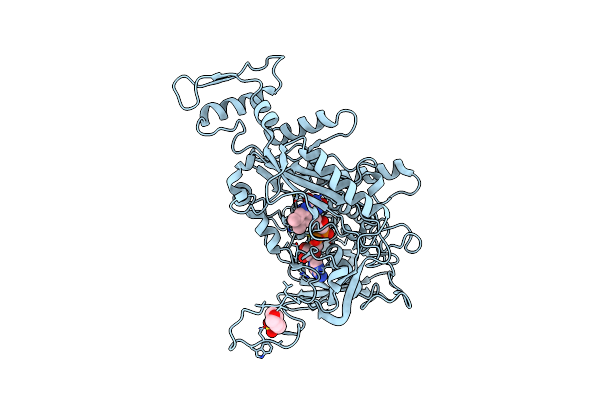
Crystal Structure Of Dutpase In Complex With Substrate Analogue Dudp And Manganese
Organism: Escherichia coli
Method: X-RAY DIFFRACTION Resolution:1.84 Å Release Date: 2007-07-31 Classification: HYDROLASE Ligands: MN, DUD, EDO |
 |
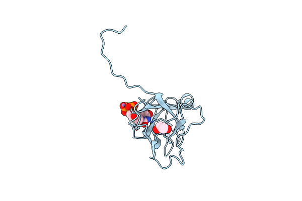
Crystal Structure Of Dutpase Complexed With Substrate Analogue Methylene-Dutp
Organism: Escherichia coli
Method: X-RAY DIFFRACTION Resolution:1.70 Å Release Date: 2007-07-31 Classification: HYDROLASE Ligands: UC5, EDO |
 |
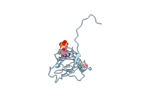
Organism: Homo sapiens
Method: X-RAY DIFFRACTION Resolution:2.20 Å Release Date: 2007-07-24 Classification: HYDROLASE Ligands: MG, DUP, CL |
 |
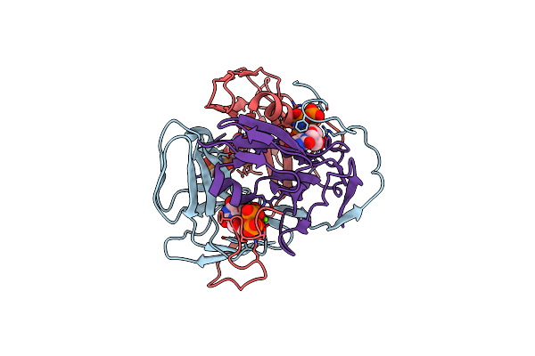
Full Length Structure Of The Mycobacterium Tuberculosis Dutpase Complexed With Magnesium And Alpha,Beta-Imido-Dutp.
Organism: Mycobacterium tuberculosis
Method: X-RAY DIFFRACTION Resolution:1.49 Å Release Date: 2007-05-22 Classification: HYDROLASE Ligands: MG, DUP, TRS |