
Search Count: 38
 |
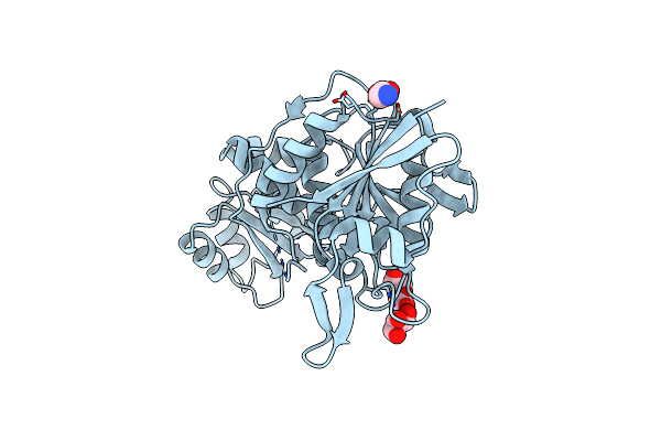
Organism: Thermococcus sibiricus
Method: X-RAY DIFFRACTION Release Date: 2025-06-11 Classification: HYDROLASE Ligands: GLY |
 |
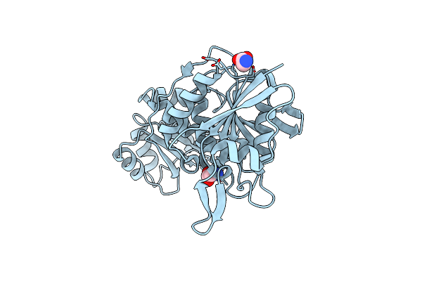
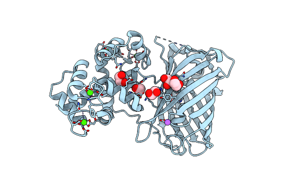
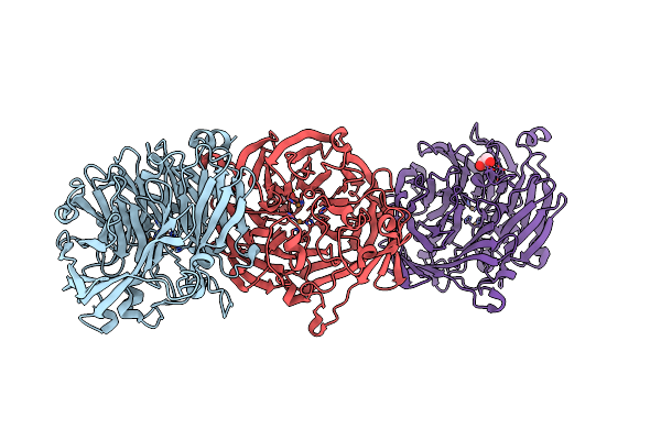
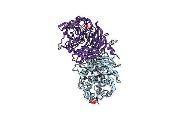
Crystal Structure Of L-Asparaginase From Thermococcus Sibiricus Double Mutation D54G/T56Q
Organism: Thermococcus sibiricus
Method: X-RAY DIFFRACTION Release Date: 2025-06-11 Classification: HYDROLASE Ligands: PEG, GOL, GLY |
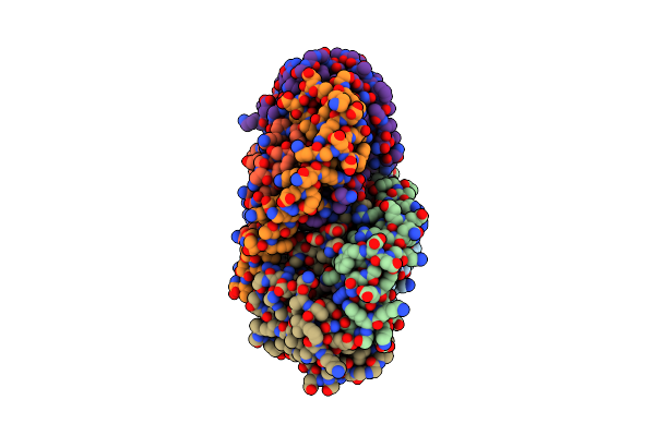 |
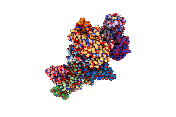
Crystal Structure Of The Soluble Green Pigment Protein From Tettigonia Cantans
Organism: Tettigonia cantans
Method: X-RAY DIFFRACTION Release Date: 2025-04-23 Classification: TRANSPORT PROTEIN Ligands: LUT, AZI, PLC, A1L6M |
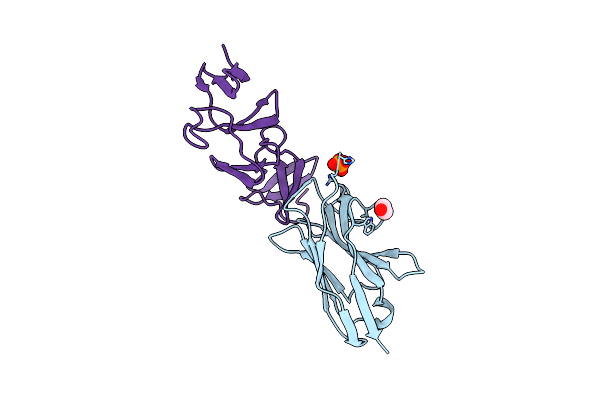 |
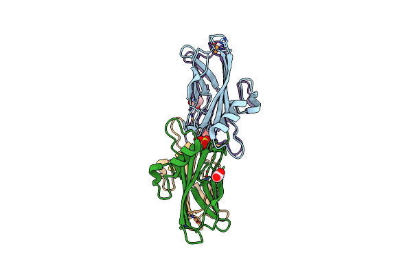
Crystal Structure Of The C. Difficile Toxin A Crops Domain Fragment 2592-2710 Bound To H5.2 Nanobody
Organism: Camelus dromedarius, Clostridioides difficile 342
Method: X-RAY DIFFRACTION Resolution:1.65 Å Release Date: 2024-12-18 Classification: TOXIN/IMMUNE SYSTEM Ligands: EDO, PO4 |
 |
Crystal Structure Of The C. Difficile Toxin A Crops Domain Fragment 2639-2707 Bound To C4.2 Nanobody
Organism: Camelus dromedarius, Clostridioides difficile
Method: X-RAY DIFFRACTION Resolution:2.10 Å Release Date: 2024-12-18 Classification: TOXIN/IMMUNE SYSTEM Ligands: EDO |
 |
Crystal Structure Of The Calcium Indicator Gcamp6S-Brus In Calcium-Bound State
Organism: Synthetic construct, Homo sapiens
Method: X-RAY DIFFRACTION Resolution:2.65 Å Release Date: 2024-12-11 Classification: FLUORESCENT PROTEIN Ligands: CA |
 |
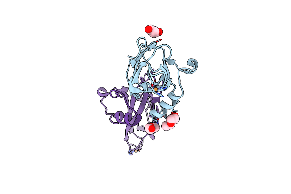
Crystal Structure Of The Calcium Indicator Gcamp6S-Brus-145 In Calcium-Bounded State
Organism: Synthetic construct, Homo sapiens
Method: X-RAY DIFFRACTION Resolution:2.05 Å Release Date: 2024-12-11 Classification: FLUORESCENT PROTEIN Ligands: EDO, CA, NA |
 |
Crystal Structure Of Beta-Carotene-Binding Protein (Bbp) From Schistocerca Gregaria Complexed With Beta-Carotene
Organism: Schistocerca gregaria
Method: X-RAY DIFFRACTION Resolution:2.70 Å Release Date: 2024-09-18 Classification: LIPID BINDING PROTEIN Ligands: BCR |
 |
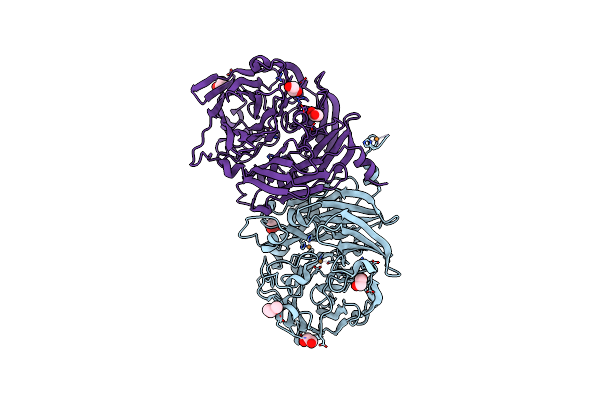
Crystal Structure Of The Complex Of Wuhan Sars-Cov-2 Rbd (319-541) With P2C5 Nanobody
Organism: Severe acute respiratory syndrome coronavirus 2, Camelus bactrianus
Method: X-RAY DIFFRACTION Resolution:3.10 Å Release Date: 2024-09-04 Classification: VIRAL PROTEIN/IMMUNE SYSTEM |
 |
Crystal Structure Of The Wuhan Sars-Cov-2 Rbd (333-541) Complexed With P2C5 Nanobody
Organism: Severe acute respiratory syndrome coronavirus 2, Camelus bactrianus
Method: X-RAY DIFFRACTION Resolution:3.70 Å Release Date: 2024-09-04 Classification: VIRAL PROTEIN/IMMUNE SYSTEM Ligands: NAG |
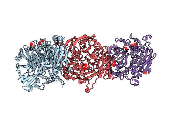 |
The Structure Of Non-Activated Thiocyanate Dehydrogenase From Pelomicrobium Methylotrophicum (Pmtcdh)
Organism: Pelomicrobium methylotrophicum
Method: X-RAY DIFFRACTION Resolution:1.45 Å Release Date: 2024-05-08 Classification: OXIDOREDUCTASE Ligands: EDO, CL, CU, PEG |
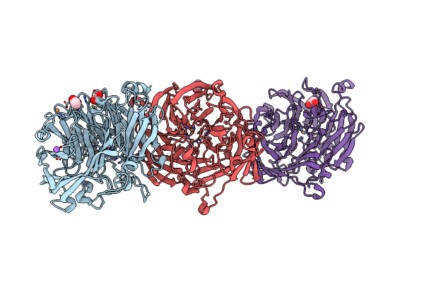 |
The Structure Of Thiocyanate Dehydrogenase From Pelomicrobium Methylotrophicum (Pmtcdh), Activated By Crystals Soaking With 1 Mm Cucl2 During 6 Months
Organism: Pelomicrobium methylotrophicum
Method: X-RAY DIFFRACTION Resolution:1.80 Å Release Date: 2024-05-08 Classification: OXIDOREDUCTASE Ligands: EDO, CU, NA |
 |
The Structure Of Thiocyanate Dehydrogenase From Pelomicrobium Methylotrophicum (Pmtcdh), Activated By Crystals Soaking With 1 Mm Cucl2 And Na Ascorbate During 12 Hours
Organism: Pelomicrobium methylotrophicum
Method: X-RAY DIFFRACTION Resolution:2.00 Å Release Date: 2024-05-08 Classification: OXIDOREDUCTASE Ligands: CU, EDO |
 |
Organism: Thioalkalivibrio paradoxus arh 1
Method: X-RAY DIFFRACTION Resolution:1.70 Å Release Date: 2024-04-24 Classification: METAL BINDING PROTEIN Ligands: CU, ACT, SO4 |
 |
Organism: Thioalkalivibrio paradoxus arh 1
Method: X-RAY DIFFRACTION Resolution:1.80 Å Release Date: 2024-04-24 Classification: METAL BINDING PROTEIN Ligands: PEG, CU |
 |
The Structure Of The Cytochrome C546/556 From Thioalkalivibrio Paradoxus With Unusual Uv-Vis Spectral Features At Atomic Resolution
Organism: Thioalkalivibrio paradoxus arh 1
Method: X-RAY DIFFRACTION Release Date: 2024-04-24 Classification: ELECTRON TRANSPORT Ligands: HEC |
 |
The Structure Of Non-Activated Thiocyanate Dehydrogenase Mutant With The H447Q Substitution From Pelomicrobium Methylotrophicum (Pmtcdh H447Q)
Organism: Pelomicrobium methylotrophicum
Method: X-RAY DIFFRACTION Resolution:1.45 Å Release Date: 2024-04-24 Classification: OXIDOREDUCTASE Ligands: PEG, EDO, CU, CL |
 |
The Structure Of Thiocyanate Dehydrogenase Mutant With The H447Q Substitution From Pelomicrobium Methylotrophicum (Pmtcdh H447Q), Activated By Crystal Soaking With 1Mm Cucl2 And 1 Mm Sodium Ascorbate
Organism: Pelomicrobium methylotrophicum
Method: X-RAY DIFFRACTION Resolution:1.55 Å Release Date: 2024-04-24 Classification: OXIDOREDUCTASE Ligands: EDO, CU, CL |
 |
Organism: Pelomicrobium methylotrophicum
Method: X-RAY DIFFRACTION Resolution:1.80 Å Release Date: 2024-03-27 Classification: OXIDOREDUCTASE Ligands: SEK, CU, GOL, SE, NA |
 |
Crystal Structure Of The Biphotochromic Fluorescent Protein Saasoti (C21N/V127T Variant) In Its Green On-State
Organism: Stylocoeniella armata
Method: X-RAY DIFFRACTION Resolution:3.00 Å Release Date: 2024-01-17 Classification: FLUORESCENT PROTEIN |