Planned Maintenance: Some services may turn out to be unavailable from 15th January, 2026 to 16th January, 2026. We apologize for the inconvenience!

Planned Maintenance: Some services may turn out to be unavailable from 15th January, 2026 to 16th January, 2026. We apologize for the inconvenience!
 |
Crystal Structure Of Wildtype Dystroglycan Proteolytic Domain (Juxtamembrane Domain)
Organism: Homo sapiens
Method: X-RAY DIFFRACTION Resolution:2.43 Å Release Date: 2024-09-11 Classification: CELL ADHESION Ligands: CA, CL |
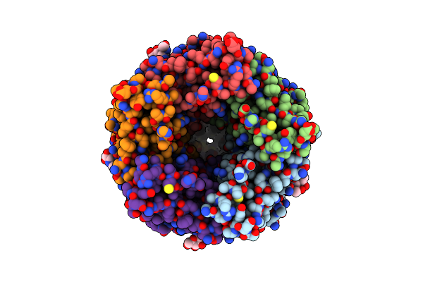 |
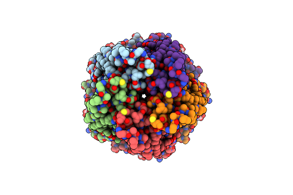
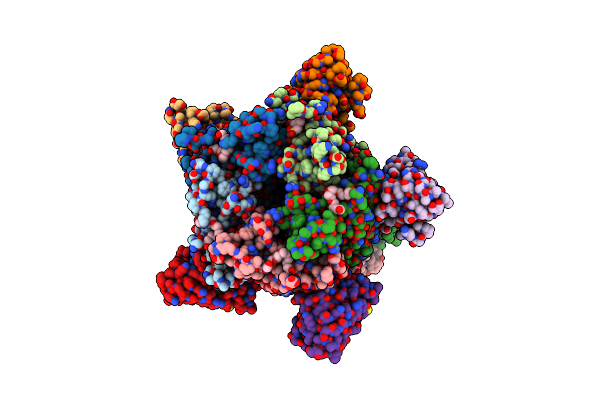
Alvinella Pompejana Nicotinic Acetylcholine Receptor Alpo4 In Apo State (Dataset 1)
Organism: Alvinella pompejana
Method: ELECTRON MICROSCOPY Release Date: 2023-07-12 Classification: MEMBRANE PROTEIN Ligands: NAG |
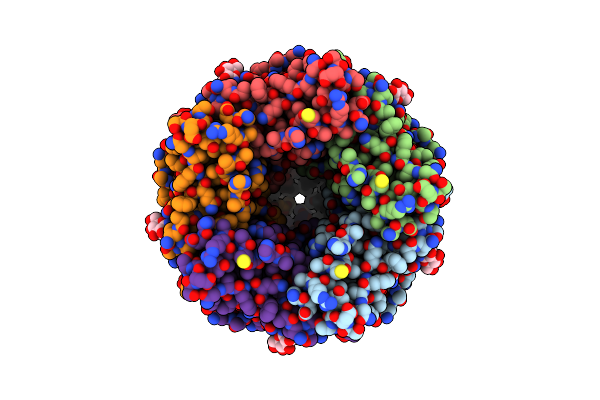 |
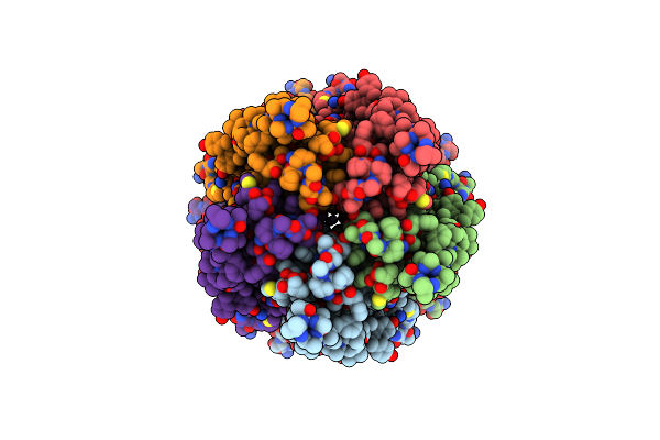
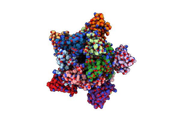
Alvinella Pompejana Nicotinic Acetylcholine Receptor Alpo In Apo State (Dataset 2)
Organism: Alvinella pompejana
Method: ELECTRON MICROSCOPY Release Date: 2023-07-12 Classification: MEMBRANE PROTEIN Ligands: NAG |
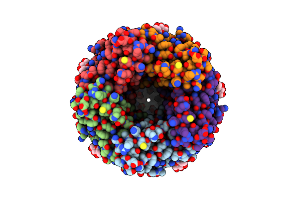 |
Alvinella Pompejana Nicotinic Acetylcholine Receptor Alpo4 In Apo State (Alpo4_Lmng_Serotonin Dataset 4)
Organism: Alvinella pompejana
Method: ELECTRON MICROSCOPY Release Date: 2023-07-12 Classification: MEMBRANE PROTEIN Ligands: NAG |
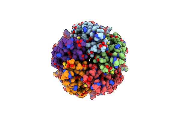 |
Alvinella Pompejana Nicotinic Acetylcholine Receptor Alpo4 In Apo State (Alpo4_Comb Dataset 3)
Organism: Alvinella pompejana
Method: ELECTRON MICROSCOPY Release Date: 2023-07-12 Classification: MEMBRANE PROTEIN Ligands: NAG |
 |
Alvinella Pompejana Nicotinic Acetylcholine Receptor Alpo4 In Apo State (Alpo4_Apo, Dataset 1)
Organism: Alvinella pompejana
Method: ELECTRON MICROSCOPY Release Date: 2023-07-12 Classification: MEMBRANE PROTEIN Ligands: NAG |
 |
Alvinella Pompejana Nicotinic Acetylcholine Receptor Alpo4 In Complex With Chaps(Alpo4_Chaps)
Organism: Alvinella pompejana
Method: ELECTRON MICROSCOPY Release Date: 2023-07-12 Classification: MEMBRANE PROTEIN Ligands: NAG, CPS, NA |
 |
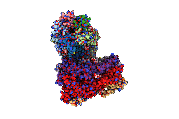
Structure Of Superoxide Dismutase 1 (Sod1) In Complex With Nanobody 2 (Nb2).
Organism: Homo sapiens, Camelus dromedarius
Method: X-RAY DIFFRACTION Resolution:2.19 Å Release Date: 2022-09-28 Classification: METAL BINDING PROTEIN Ligands: CU, ZN |
 |
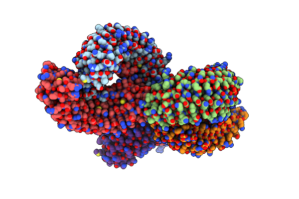
Cryo-Em Structure Of Torpedo Nicotinic Acetylcholine Receptor In Complex With A Short-Chain Neurotoxin.
Organism: Synthetic construct, Tetronarce californica
Method: ELECTRON MICROSCOPY Release Date: 2022-08-17 Classification: MEMBRANE PROTEIN Ligands: NAG |
 |
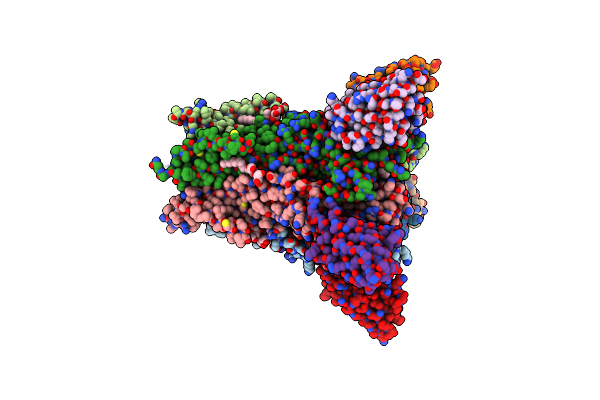
The Crystal Structure Of A Trp Channel Peptide Bound To A G Protein Beta Gamma Heterodimer
Organism: Mus musculus
Method: X-RAY DIFFRACTION Resolution:1.94 Å Release Date: 2020-10-14 Classification: PROTEIN BINDING Ligands: GOL |
 |
Organism: Homo sapiens
Method: X-RAY DIFFRACTION Resolution:1.67 Å Release Date: 2020-03-11 Classification: CELL CYCLE Ligands: GOL, ZN |
 |
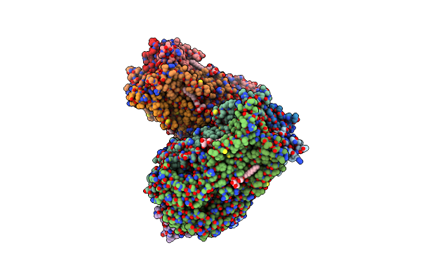
Structure Of The Pentameric Ligand-Gated Ion Channel Elic In Complex With A Pam Nanobody
Organism: Dickeya chrysanthemi, Lama glama
Method: X-RAY DIFFRACTION Resolution:2.59 Å Release Date: 2020-02-12 Classification: TRANSPORT PROTEIN Ligands: ABU, MES, GOL, CA, PG4, UMQ, ACT |
 |
Structure Of The Pentameric Ligand-Gated Ion Channel Elic In Complex With A Nam Nanobody
Organism: Dickeya chrysanthemi, Lama glama
Method: X-RAY DIFFRACTION Resolution:3.25 Å Release Date: 2020-02-12 Classification: TRANSPORT PROTEIN Ligands: CA, UMQ |
 |
X-Ray Structure Of A Pentameric Ligand Gated Ion Channel From Erwinia Chrysanthemi (Elic) 7'C Pore Mutant (L238C) In Complex With Nanobody 72
Organism: Dickeya chrysanthemi, Lama glama
Method: X-RAY DIFFRACTION Resolution:2.50 Å Release Date: 2019-10-09 Classification: MEMBRANE PROTEIN Ligands: P6G, PTY, LMT, MES, NA |
 |
X-Ray Structure Of A Pentameric Ligand Gated Ion Channel From Erwinia Chrysanthemi (Elic) Delta8 Truncation Mutant In Complex With Nanobody 72
Organism: Dickeya chrysanthemi, Lama glama
Method: X-RAY DIFFRACTION Resolution:2.78 Å Release Date: 2019-10-09 Classification: MEMBRANE PROTEIN |
 |
X-Ray Structure Of A Pentameric Ligand Gated Ion Channel From Erwinia Chrysanthemi (Elic) F16'S Pore Mutant (F247S) With Alternate M4 Conformation.
Organism: Dickeya chrysanthemi
Method: X-RAY DIFFRACTION Resolution:3.45 Å Release Date: 2019-10-09 Classification: MEMBRANE PROTEIN Ligands: LMT |
 |
Organism: Homo sapiens
Method: X-RAY DIFFRACTION Resolution:3.09 Å Release Date: 2019-01-30 Classification: SIGNALING PROTEIN Ligands: SO4 |
 |
Humanized Alpha-Achbp (Acetylcholine Binding Protein) In Complex With Lobeline And Allosteric Binder Fragment 4.
Organism: Homo sapiens
Method: X-RAY DIFFRACTION Resolution:2.57 Å Release Date: 2017-12-20 Classification: CHOLINE-BINDING PROTEIN Ligands: L0B, 9Z0, DMS |
 |
Humanized Alpha-Achbp (Acetylcholine Binding Protein) In Complex With Lobeline.
Organism: Homo sapiens
Method: X-RAY DIFFRACTION Resolution:2.50 Å Release Date: 2017-11-29 Classification: CHOLINE-BINDING PROTEIN Ligands: DMS, L0B |
 |
Humanized Alpha-Achbp (Acetylcholine Binding Protein) In Complex With Allosteric Binder Fragment Cu2017
Organism: Homo sapiens, Lymnaea stagnalis
Method: X-RAY DIFFRACTION Resolution:3.10 Å Release Date: 2017-11-29 Classification: CHOLINE-BINDING PROTEIN Ligands: AVB, EDO, PEG |