
Search Count: 408
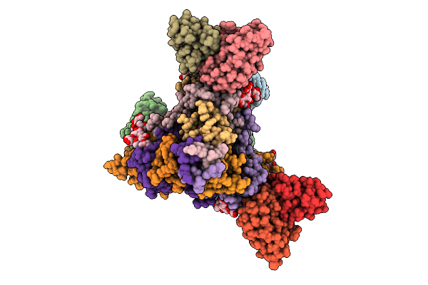 |
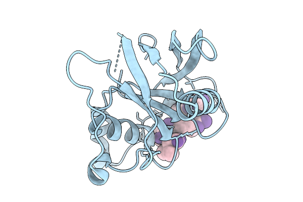
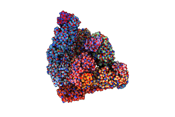
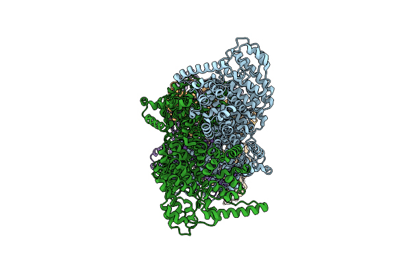
A Broadly-Neutralizing Antibody Against Ebolavirus Glycoprotein That Can Potentiate The Breadth And Neutralization Potency Of Other Anti-Glycoprotein Antibodies
Organism: Oryctolagus cuniculus, Ebolavirus
Method: ELECTRON MICROSCOPY Release Date: 2025-11-05 Classification: VIRAL PROTEIN/IMMUNE SYSTEM Ligands: NAG |
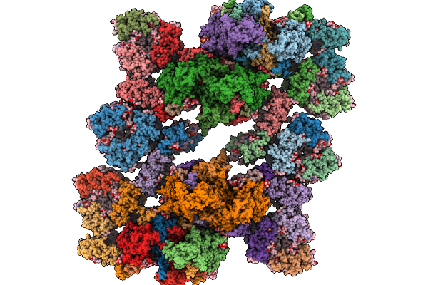 |
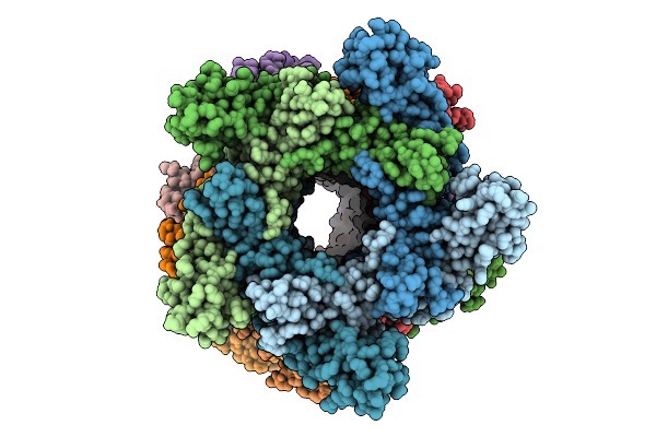
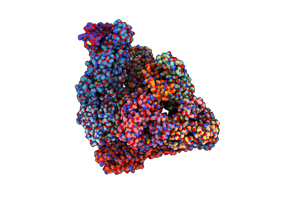
Coordinates Of Cryo-Em Structure Of The Arabidopsis Thaliana C4S4M4-Type Psii Supercomplex
Organism: Arabidopsis thaliana
Method: ELECTRON MICROSCOPY Release Date: 2025-10-08 Classification: PHOTOSYNTHESIS Ligands: CHL, CLA, LUT, NEX, LHG, XAT, BCR, LMG, PL9, SQD, HEM, DGD, BCT, OEX, FE2, PHO |
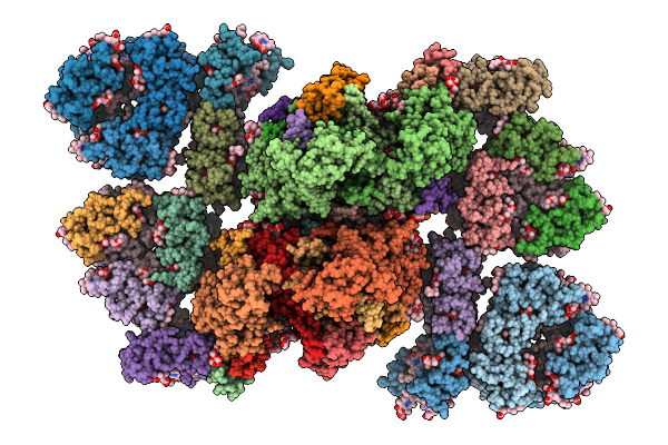 |
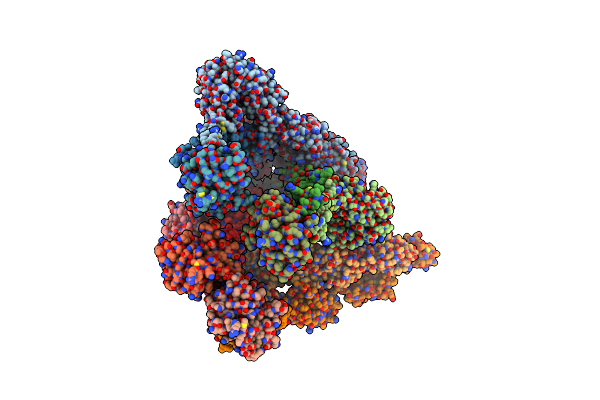
Coordinates Of Cryo-Em Structure Of The Arabidopsis Thaliana C2S2M2-Type Psii Supercomplex
Organism: Arabidopsis thaliana
Method: ELECTRON MICROSCOPY Release Date: 2025-10-08 Classification: PHOTOSYNTHESIS Ligands: CHL, CLA, LUT, NEX, XAT, LHG, BCR, OEX, FE2, PHO, LMG, SQD, DGD, BCT, PL9, HEM |
 |
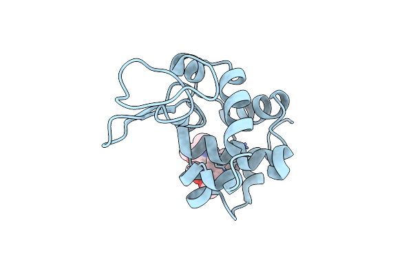
Arabidopsis Thaliana Protease-Associated Domain Of Vacuolar Sorting Receptor 1 In Complexed With Vicilin-Like Seed Storage Protein 22 C-Terminal Pentapeptide Sdrfv (Ph 5.8)
Organism: Arabidopsis thaliana
Method: X-RAY DIFFRACTION Release Date: 2025-09-10 Classification: PROTEIN TRANSPORT Ligands: TRS, GOL, PEG |
 |
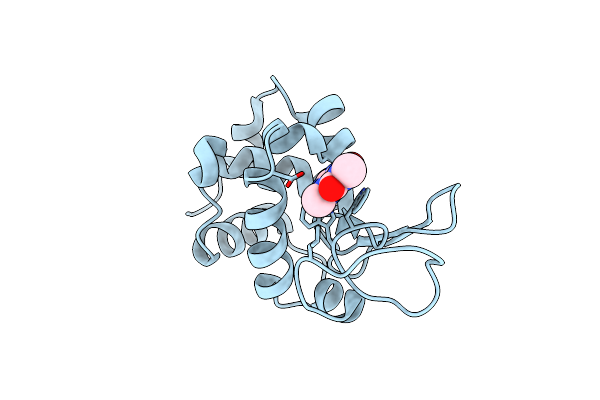
Arabidopsis Thaliana Protease-Associated Domain Of Vacuolar Sorting Receptor 1 In Complexed With Vicilin-Like Seed Storage Protein 22 C-Terminal Pentapeptide Sdrfv (Ph 7.0)
Organism: Arabidopsis thaliana
Method: X-RAY DIFFRACTION Release Date: 2025-09-10 Classification: PROTEIN TRANSPORT |
 |
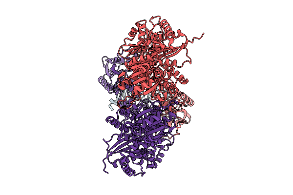
Organism: Homo sapiens
Method: ELECTRON MICROSCOPY Release Date: 2025-08-06 Classification: RNA BINDING PROTEIN Ligands: ZN |
 |
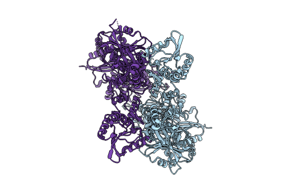
Electronic Microscopy Structure Of Human Schlafen14-E211A Dimer In Complex With Dsrna
Organism: Homo sapiens
Method: ELECTRON MICROSCOPY Release Date: 2025-08-06 Classification: RNA BINDING PROTEIN/RNA Ligands: ZN |
 |
Organism: Bacillus subtilis
Method: ELECTRON MICROSCOPY Release Date: 2025-07-16 Classification: ANTIVIRAL PROTEIN |
 |
Organism: Bacillus subtilis
Method: ELECTRON MICROSCOPY Release Date: 2025-07-16 Classification: ANTIVIRAL PROTEIN |
 |
Organism: Homo sapiens
Method: ELECTRON MICROSCOPY Release Date: 2025-06-18 Classification: ONCOPROTEIN |
 |
Organism: Homo sapiens
Method: ELECTRON MICROSCOPY Release Date: 2025-06-18 Classification: ONCOPROTEIN/INHIBITOR |
 |
Organism: Gallus gallus
Method: X-RAY DIFFRACTION Resolution:1.50 Å Release Date: 2025-04-23 Classification: HYDROLASE Ligands: 34G |
 |
Organism: Gallus gallus
Method: X-RAY DIFFRACTION Resolution:1.48 Å Release Date: 2025-04-23 Classification: HYDROLASE Ligands: TEP |
 |
Organism: Western equine encephalitis virus, Homo sapiens
Method: ELECTRON MICROSCOPY Release Date: 2025-04-23 Classification: VIRAL PROTEIN |
 |
Organism: Western equine encephalitis virus, Homo sapiens
Method: ELECTRON MICROSCOPY Release Date: 2025-04-23 Classification: VIRAL PROTEIN Ligands: CA |
 |
Organism: Western equine encephalitis virus, Homo sapiens
Method: ELECTRON MICROSCOPY Release Date: 2025-04-23 Classification: VIRAL PROTEIN Ligands: CA |
 |
Organism: Ross river virus (strain t48), Homo sapiens
Method: ELECTRON MICROSCOPY Release Date: 2025-04-23 Classification: VIRAL PROTEIN Ligands: CA |
 |
Organism: Bacillus subtilis a29
Method: ELECTRON MICROSCOPY Release Date: 2025-03-05 Classification: HYDROLASE |
 |
Organism: Bacillus subtilis a29
Method: ELECTRON MICROSCOPY Release Date: 2025-03-05 Classification: HYDROLASE |
 |
Organism: Bacillus subtilis a29
Method: ELECTRON MICROSCOPY Release Date: 2025-03-05 Classification: HYDROLASE |