
Search Count: 12
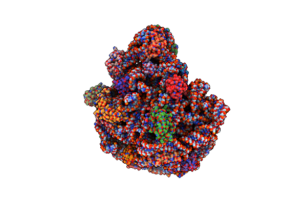 |
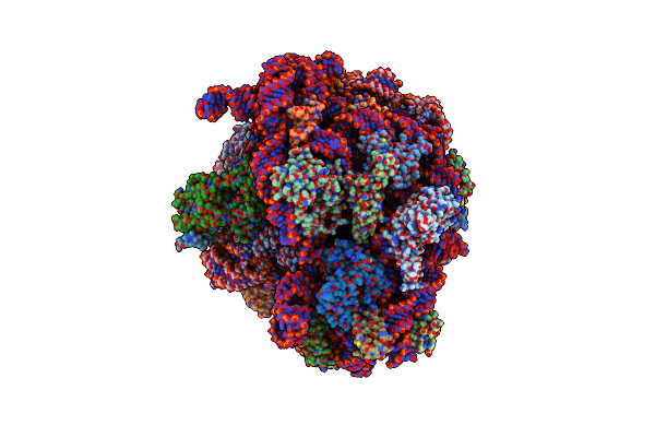
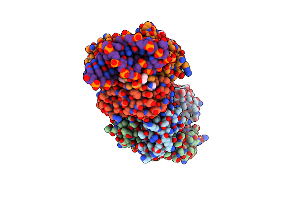
Structure Of The 50S Ribosomal Subunit From The Antibiotic-Producing Bacterium Streptomyces Fradiae
Organism: Streptomyces fradiae atcc 10745 = dsm 40063
Method: ELECTRON MICROSCOPY Release Date: 2025-05-14 Classification: RIBOSOME |
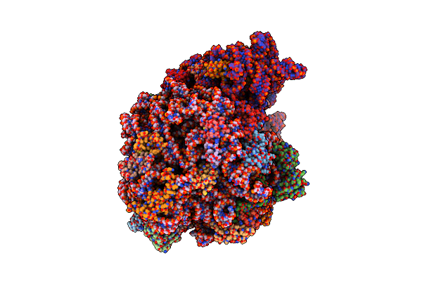 |
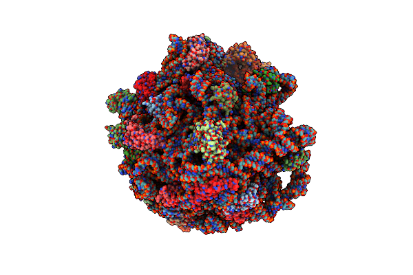
Organism: Psychrobacter urativorans
Method: ELECTRON MICROSCOPY Release Date: 2025-03-26 Classification: RIBOSOME |
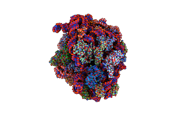 |
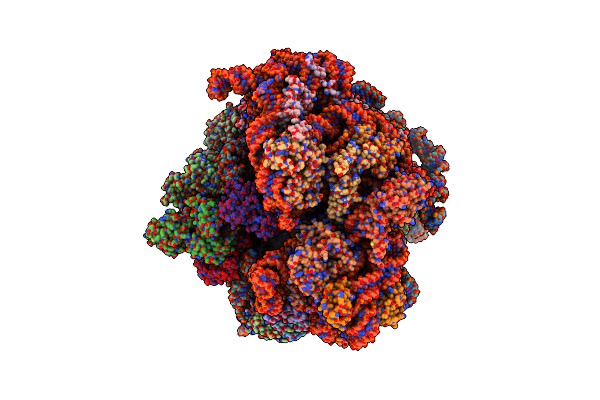
Cryo-Em Structure Of P. Urativorans 70S Ribosome In Complex With Hibernation Factors Balon And Raia (Structure 1).
Organism: Psychrobacter urativorans
Method: ELECTRON MICROSCOPY Release Date: 2024-02-21 Classification: RIBOSOME Ligands: MG |
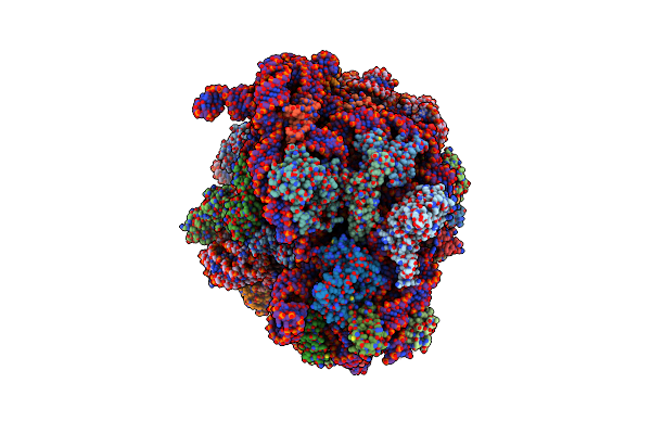 |
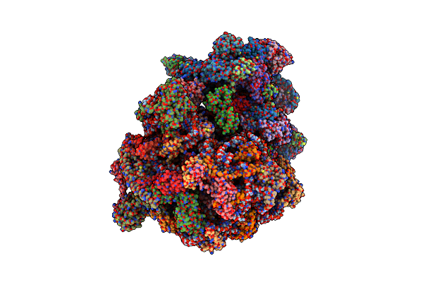
Cryo-Em Structure Of P. Urativorans 70S Ribosome In Complex With Hibernation Factor Balon, Mrna And P-Site Trna (Structure 2).
Organism: Psychrobacter urativorans
Method: ELECTRON MICROSCOPY Release Date: 2024-02-21 Classification: RIBOSOME |
 |
Cryo-Em Structure Of P. Urativorans 70S Ribosome In Complex With Hibernation Factor Balon And Ef-Tu(Gdp) (Structure 3).
Organism: Psychrobacter urativorans
Method: ELECTRON MICROSCOPY Release Date: 2024-02-21 Classification: RIBOSOME Ligands: GDP, MG |
 |
Cryo-Em Structure Of The Mycobacterium Smegmatis 70S Ribosome In Complex With Hibernation Factor Msmeg1130 (Balon) (Structure 4)
Organism: Mycolicibacterium smegmatis mc2 155
Method: ELECTRON MICROSCOPY Release Date: 2024-02-07 Classification: RIBOSOME Ligands: ZN |
 |
Cryo-Em Structure Of The Mycobacterium Smegmatis 70S Ribosome In Complex With Hibernation Factor Rv2629 (Balon) (Structure 5)
Organism: Mycobacterium tuberculosis h37rv, Mycolicibacterium smegmatis mc2 155
Method: ELECTRON MICROSCOPY Release Date: 2024-02-07 Classification: RIBOSOME Ligands: ZN |
 |
Cryo-Em Structure Of The Mycobacterium Smegmatis 70S Ribosome In Complex With Hibernation Factor Msmeg1130 (Balon) And Msmegef-Tu(Gdp) (Composite Structure 6)
Organism: Mycolicibacterium smegmatis mc2 155
Method: ELECTRON MICROSCOPY Release Date: 2024-02-07 Classification: RIBOSOME Ligands: GDP, ZN |
 |
Organism: Encephalitozoon cuniculi gb-m1
Method: ELECTRON MICROSCOPY Release Date: 2022-02-09 Classification: RIBOSOME Ligands: ZN, AMP, SPD |
 |
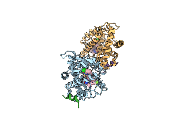
Organism: Homo sapiens, Synthetic construct
Method: X-RAY DIFFRACTION Resolution:1.70 Å Release Date: 2022-02-02 Classification: IMMUNE SYSTEM/DNA Ligands: EDO, SO4, IMD |
 |
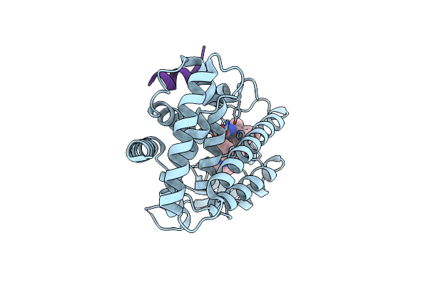
Crystal Structure Of Ppargamma Ligand Binding Domain In Complex With Dibenzooxepine Derivative Compound-17
Organism: Homo sapiens
Method: X-RAY DIFFRACTION Resolution:1.84 Å Release Date: 2019-10-30 Classification: NUCLEAR PROTEIN Ligands: CTU |
 |
Crystal Structure Of Ppargamma Ligand Binding Domain In Complex With Dibenzooxepine Derivative Compound-9
Organism: Homo sapiens
Method: X-RAY DIFFRACTION Resolution:2.20 Å Release Date: 2018-11-14 Classification: NUCLEAR PROTEIN Ligands: KK4 |