
Search Count: 22
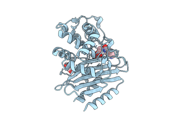 |
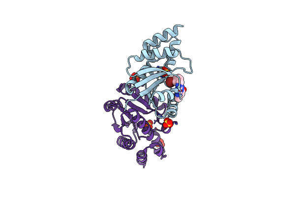
Crystal Structure Of Peni Beta-Lactamase From Burkholderia Pseudomallei Complex With Taniborbactam
Organism: Burkholderia pseudomallei k96243
Method: X-RAY DIFFRACTION Release Date: 2025-08-13 Classification: HYDROLASE Ligands: KJK, GOL |
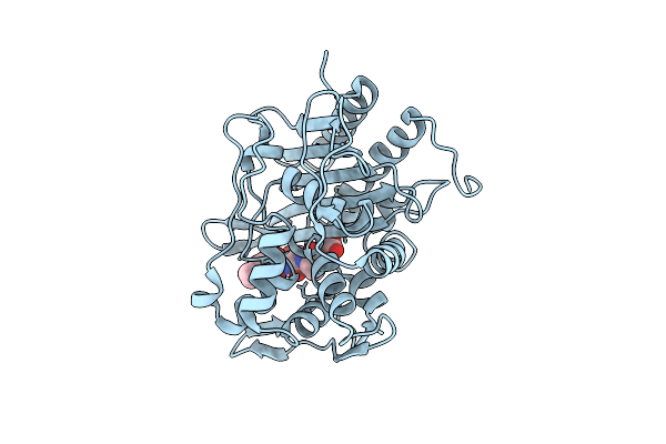 |
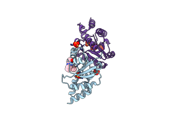
Crystal Structure Of The Transpeptidase Domain Of Pbp2 From The Neisseria Gonorrhoeae Cephalosporin Decreased Susceptibility Strain 35/02 In Complex With Boronate Inhibitor Vnrx-6884
Organism: Neisseria gonorrhoeae 35/02
Method: X-RAY DIFFRACTION Resolution:1.89 Å Release Date: 2025-01-22 Classification: LIGASE Ligands: A1BJC |
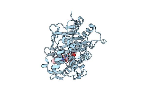 |
Crystal Structure Of The Transpeptidase Domain Of Pbp2 From The Neisseria Gonorrhoeae Cephalosporin Decreased Susceptibility Strain 35/02 In Complex With Boronate Inhibitor Vnrx-6752
Organism: Neisseria gonorrhoeae 35/02
Method: X-RAY DIFFRACTION Resolution:2.61 Å Release Date: 2025-01-22 Classification: LIGASE Ligands: A1BJB |
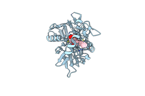 |
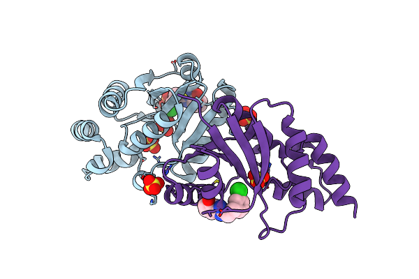
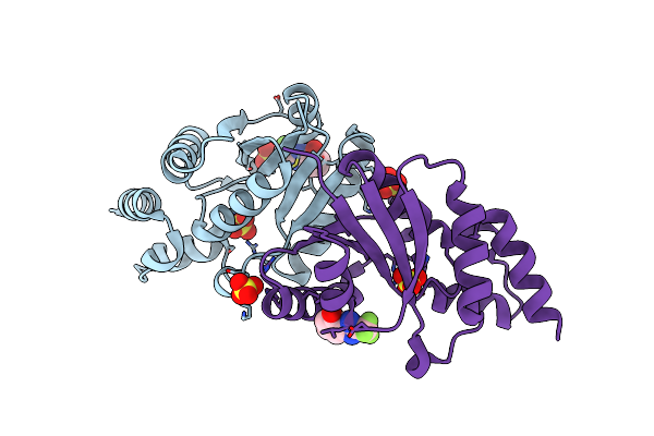
Chain A. Udp-3-O-[3-Hydroxymyristoyl] N-Acetylglucosamine Deacetylase Pa-Lpxc Complexed With (R)-3-((S)-3-(4-(Cyclopropylethynyl)Phenyl)-2-Oxooxazolidin-5-Yl)-N-Hydroxy-2-Methyl-2-(Methylsulfonyl)Propenamide
Organism: Pseudomonas aeruginosa (strain atcc 15692 / dsm 22644 / cip 104116 / jcm 14847 / lmg 12228 / 1c / prs 101 / pao1)
Method: X-RAY DIFFRACTION Resolution:1.80 Å Release Date: 2019-01-23 Classification: HYDROLASE Ligands: JBA, ZN |
 |
Phosphopantetheine Adenylyltransferase (Coad) In Complex With (R)-3-(3-Chlorophenyl)-3-((5-Methyl-7-Oxo-4,7-Dihydro-[1,2,4]Triazolo[1,5-A]Pyrimidin-2-Yl)Amino)Propanenitrile
Organism: Escherichia coli (strain k12)
Method: X-RAY DIFFRACTION Resolution:2.20 Å Release Date: 2018-04-04 Classification: TRANSFERASE/ANTIBIOTIC Ligands: EXJ, SO4, DMS |
 |
Phosphopantetheine Adenylyltransferase (Coad) In Complex With N-(2-(5-Methoxy-1H-Indol-3-Yl)Ethyl)Pivalamide
Organism: Escherichia coli (strain k12)
Method: X-RAY DIFFRACTION Resolution:2.28 Å Release Date: 2018-04-04 Classification: TRANSFERASE/ANTIBIOTIC Ligands: F1V, SO4 |
 |
Phosphopantetheine Adenylyltransferase (Coad) In Complex With Methyl (R)-4-(3-(2-Cyano-1-((5-Methyl-7-Oxo-4,7-Dihydro-[1,2,4]Triazolo[1,5-A]Pyrimidin-2-Yl)Amino)Ethyl)Phenoxy)Piperidine-1-Carboxylate
Organism: Escherichia coli (strain k12)
Method: X-RAY DIFFRACTION Resolution:2.03 Å Release Date: 2018-04-04 Classification: TRANSFERASE/ANTIBIOTIC Ligands: F1D, SO4, PEG, PG4 |
 |
Phosphopantetheine Adenylyltransferase (Coad) In Complex With (R)-2-((1-(3-(4-Methoxyphenoxy)Phenyl)Ethyl)Amino)-5-Methyl-[1,2,4]Triazolo[1,5-A]Pyrimidin-7(4H)-One
Organism: Escherichia coli (strain k12)
Method: X-RAY DIFFRACTION Resolution:1.85 Å Release Date: 2018-04-04 Classification: TRANSFERASE/ANTIBIOTIC Ligands: F14, SO4, PG4 |
 |
Phosphopantetheine Adenylyltransferase (Coad) In Complex With Methyl (R)-4-(3-(2-Cyano-1-((5-Methyl-1H-Imidazo[4,5-B]Pyridin-2-Yl)Amino)Ethyl)Benzyl)Piperidine-1-Carboxylate
Organism: Escherichia coli (strain k12)
Method: X-RAY DIFFRACTION Resolution:1.94 Å Release Date: 2018-04-04 Classification: TRANSFERASE/ANTIBIOTIC Ligands: F0Y, SO4, PG4 |
 |
Phosphopantetheine Adenylyltransferase (Coad) In Complex With 2-Benzyl-N-(3-Chloro-4-Methylphenyl)-5-Methyl-[1,2,4]Triazolo[1,5-A]Pyrimidin-7-Amine
Organism: Escherichia coli (strain k12)
Method: X-RAY DIFFRACTION Resolution:1.79 Å Release Date: 2018-04-04 Classification: TRANSFERASE/ANTIBIOTIC Ligands: F0V, SO4, DMS, PG4 |
 |
Phosphopantetheine Adenylyltransferase (Coad) In Complex With (R)-3-((7-(((S)-2-Amino-2-(2-Methoxyphenyl)Ethyl)Amino)-5-Methyl-[1,2,4]Triazolo[1,5-A]Pyrimidin-2-Yl)Amino)-3-(3-Chlorophenyl)Propanenitrile
Organism: Escherichia coli
Method: X-RAY DIFFRACTION Resolution:2.06 Å Release Date: 2018-04-04 Classification: TRANSFERASE/ANTIBIOTIC Ligands: F6D, SO4, DMS, PG4 |
 |
Crystal Structure Of E.Coli Phosphopantetheine Adenylyltransferase (Ppat/Coad) In Complex With (R)-3-(3-Chlorophenyl)-3-((5-Methyl-7-Oxo-4,7-Dihydro-[1,2,4]Triazolo[1,5-A]Pyrimidin-2-Yl)Amino)Propanenitrile
Organism: Escherichia coli (strain k12)
Method: X-RAY DIFFRACTION Resolution:1.61 Å Release Date: 2018-03-14 Classification: TRANSFERASE/antibiotic Ligands: EXJ, ATP, MG, SO4 |
 |
Crystal Structure Of E.Coli Phosphopantetheine Adenylyltransferase (Ppat/Coad) In Complex With 1-Benzyl-1H-Imidazo[4,5-B]Pyridine
Organism: Escherichia coli (strain k12)
Method: X-RAY DIFFRACTION Resolution:1.77 Å Release Date: 2018-03-14 Classification: TRANSFERASE/antibiotic Ligands: EXG, SO4, DMS |
 |
Crystal Structure Of E.Coli Phosphopantetheine Adenylyltransferase (Ppat/Coad) In Complex With 2-((3-Bromobenzyl)Amino)-5-Methyl-[1,2,4]Triazolo[1,5-A]Pyrimidin-7(4H)-One
Organism: Escherichia coli (strain k12)
Method: X-RAY DIFFRACTION Resolution:1.79 Å Release Date: 2018-03-14 Classification: TRANSFERASE/antibiotic Ligands: EXP, SO4 |
 |
Crystal Structure Of E.Coli Phosphopantetheine Adenylyltransferase (Ppat/Coad) In Complex With (R)-2,4-Dihydroxy-N-(2-(4-Hydroxy-1H-Benzo[D]Imidazol-2-Yl)Ethyl)-3,3-Dimethylbutanamide
Organism: Escherichia coli (strain k12)
Method: X-RAY DIFFRACTION Resolution:1.87 Å Release Date: 2018-03-14 Classification: TRANSFERASE/antibiotic Ligands: EXS, SO4 |
 |
Crystal Structure Of E.Coli Phosphopantetheine Adenylyltransferase (Ppat/Coad) In Complex With 3-((1S,2S)-2-(4-Hydroxy-1H-Benzo[D]Imidazol-2-Yl)Cyclopentyl)Benzoic Acid
Organism: Escherichia coli (strain k12)
Method: X-RAY DIFFRACTION Resolution:1.82 Å Release Date: 2018-03-14 Classification: TRANSFERASE/antibiotic Ligands: EXV, SO4 |
 |
Crystal Structure Of E.Coli Phosphopantetheine Adenylyltransferase (Ppat/Coad) In Complex With 2-(3-Chlorophenethyl)-1H-Benzo[D]Imidazol-4-Ol
Organism: Escherichia coli (strain k12)
Method: X-RAY DIFFRACTION Resolution:1.92 Å Release Date: 2018-03-14 Classification: TRANSFERASE/antibiotic Ligands: EX7, SO4 |
 |
Crystal Structure Of E.Coli Phosphopantetheine Adenylyltransferase (Ppat/Coad) In Complex With 2-(Trifluoromethyl)-1H-Benzo[D]Imidazol-4-Ol
Organism: Escherichia coli (strain k12)
Method: X-RAY DIFFRACTION Resolution:2.06 Å Release Date: 2018-03-14 Classification: TRANSFERASE/antibiotic Ligands: EXD, SO4 |
 |
Organism: Influenza a virus (strain a/udorn/307/1972 h3n2)
Method: X-RAY DIFFRACTION Resolution:2.40 Å Release Date: 2017-09-06 Classification: VIRAL PROTEIN/INHIBITOR Ligands: 21G, PEG |
 |
Organism: Influenza b virus
Method: X-RAY DIFFRACTION Resolution:1.70 Å Release Date: 2015-11-18 Classification: PROTEIN BINDING |