
Search Count: 71
 |
Organism: Vibrio cholerae
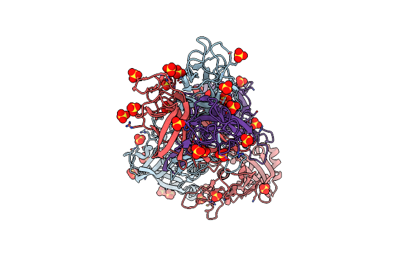
Method: X-RAY DIFFRACTION Resolution:2.32 Å Release Date: 2022-11-09 Classification: CELL ADHESION Ligands: SO4 |
 |
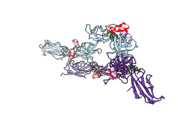
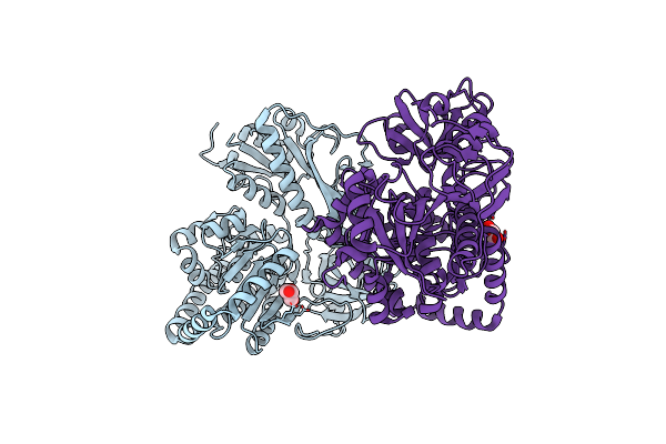
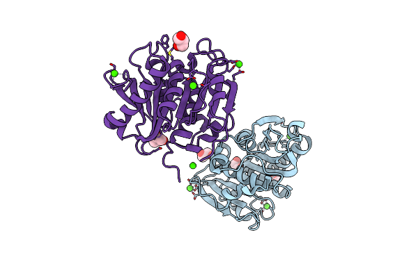
Crystal Structure Of Minor Pilin Tcpb From Vibrio Cholerae Complexed With N-Terminal Peptide Fragment Of Tcpf
Organism: Vibrio cholerae
Method: X-RAY DIFFRACTION Resolution:2.30 Å Release Date: 2022-11-09 Classification: CELL ADHESION Ligands: CA, CL, 1PE |
 |
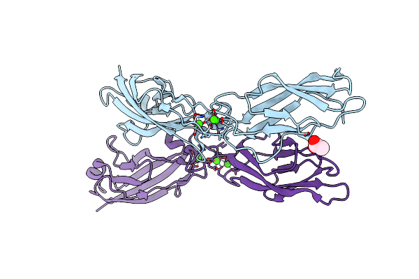
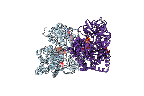
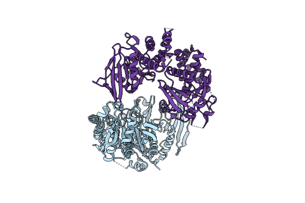
Crystal Structure Of Minor Pilin Tcpb From Vibrio Cholerae Complexed With Secreted Protein Tcpf
Organism: Vibrio cholerae
Method: X-RAY DIFFRACTION Resolution:4.05 Å Release Date: 2022-11-09 Classification: CELL ADHESION |
 |
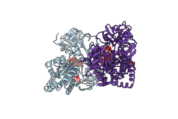
Organism: Homo sapiens
Method: X-RAY DIFFRACTION Resolution:2.70 Å Release Date: 2021-08-18 Classification: CELL ADHESION Ligands: NAG, CA |
 |
Organism: Homo sapiens
Method: X-RAY DIFFRACTION Resolution:1.38 Å Release Date: 2021-06-16 Classification: CELL ADHESION Ligands: CA, ACT, NA |
 |
Organism: Synthetic construct
Method: X-RAY DIFFRACTION Resolution:1.75 Å Release Date: 2020-11-25 Classification: RNA/NUCLEIC ACID |
 |
Organism: Synthetic construct
Method: X-RAY DIFFRACTION Resolution:1.70 Å Release Date: 2020-11-25 Classification: RNA/NUCLEIC ACID Ligands: CA |
 |
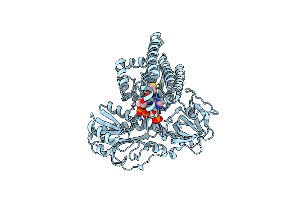
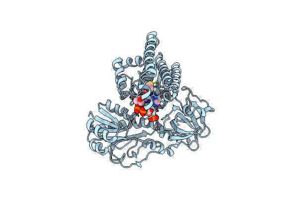
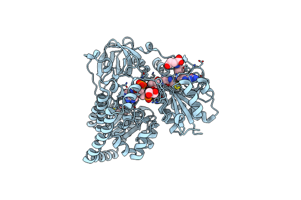
Crystal Structure Of Gmp Reductase From Trypanosoma Brucei In Complex With Guanosine 5'-Triphosphate
Organism: Trypanosoma brucei brucei
Method: X-RAY DIFFRACTION Resolution:2.50 Å Release Date: 2020-03-18 Classification: OXIDOREDUCTASE Ligands: GTP, PO4 |
 |
Organism: Trypanosoma brucei brucei (strain iltat1.4)
Method: X-RAY DIFFRACTION Resolution:2.80 Å Release Date: 2020-03-04 Classification: OXIDOREDUCTASE |
 |
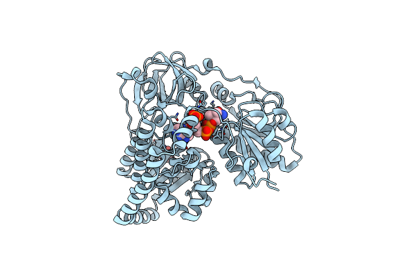
Crystal Structure Of Gmp Reductase C318A From Trypanosoma Brucei In Complex With Guanosine 5'-Monophosphate
Organism: Trypanosoma brucei brucei (strain iltat1.4)
Method: X-RAY DIFFRACTION Resolution:1.90 Å Release Date: 2020-02-26 Classification: OXIDOREDUCTASE Ligands: 5GP, K |
 |
Organism: Aquifex aeolicus (strain vf5)
Method: X-RAY DIFFRACTION Resolution:1.79 Å Release Date: 2019-11-06 Classification: BIOSYNTHETIC PROTEIN Ligands: COA, GOL |
 |
Organism: Aquifex aeolicus vf5
Method: X-RAY DIFFRACTION Resolution:2.40 Å Release Date: 2019-11-06 Classification: BIOSYNTHETIC PROTEIN Ligands: COA |
 |
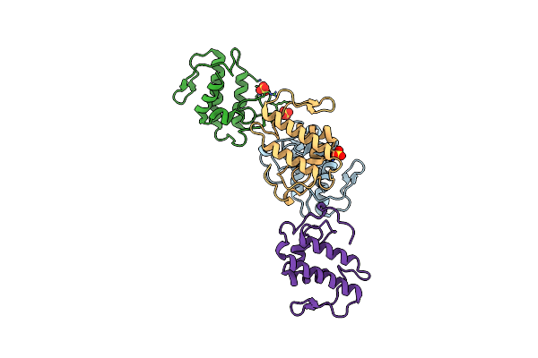
Crystal Structure Of Hypx From Aquifex Aeolicus In Complex With Tetrahydrofolic Acid
Organism: Aquifex aeolicus (strain vf5)
Method: X-RAY DIFFRACTION Resolution:2.10 Å Release Date: 2019-11-06 Classification: BIOSYNTHETIC PROTEIN Ligands: COA, THG, GOL |
 |
Crystal Structure Of Hypx From Aquifex Aeolicus, R9A-Q15A-R131A-R542A Variant
Organism: Aquifex aeolicus vf5
Method: X-RAY DIFFRACTION Resolution:2.50 Å Release Date: 2019-11-06 Classification: BIOSYNTHETIC PROTEIN Ligands: COA, GOL |
 |
Crystal Structure Of Hypx From Aquifex Aeolicus, Q15A-R131A-S194A-Q195A-N306A-R542A Variant
Organism: Aquifex aeolicus (strain vf5)
Method: X-RAY DIFFRACTION Resolution:2.10 Å Release Date: 2019-11-06 Classification: BIOSYNTHETIC PROTEIN Ligands: GOL |
 |
Organism: Aquifex aeolicus vf5
Method: X-RAY DIFFRACTION Resolution:2.29 Å Release Date: 2019-11-06 Classification: BIOSYNTHETIC PROTEIN Ligands: COA, GOL |
 |
Crystal Structure Of Hypx From Aquifex Aeolicus, A392F-I419F Variant In Complex With Tetrahydrofolic Acid
Organism: Aquifex aeolicus vf5
Method: X-RAY DIFFRACTION Resolution:2.00 Å Release Date: 2019-11-06 Classification: BIOSYNTHETIC PROTEIN Ligands: COA, THG, GOL |
 |
Organism: Rattus norvegicus
Method: X-RAY DIFFRACTION Resolution:3.32 Å Release Date: 2019-08-28 Classification: HYDROLASE |
 |
Organism: Protobothrops flavoviridis
Method: X-RAY DIFFRACTION Resolution:2.57 Å Release Date: 2019-01-16 Classification: TOXIN Ligands: SO4 |
 |
Crystal Structure Of Pet-Degrading Cutinase Cut190 S176A/S226P/R228S/ Mutant In Ca(2+)-Bound State
Organism: Saccharomonospora viridis
Method: X-RAY DIFFRACTION Resolution:1.60 Å Release Date: 2018-09-12 Classification: HYDROLASE Ligands: CA, GOL |