
Search Count: 16
 |
Organism: Clostridium perfringens
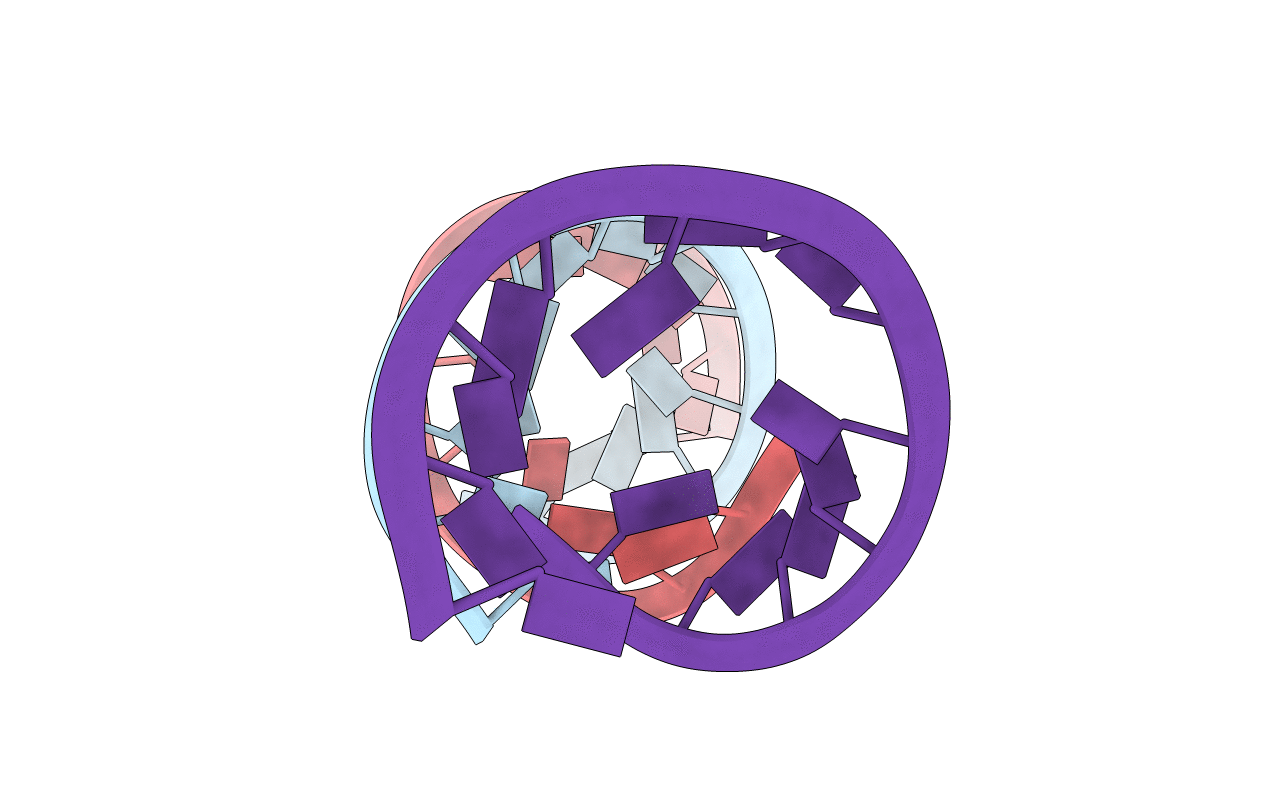
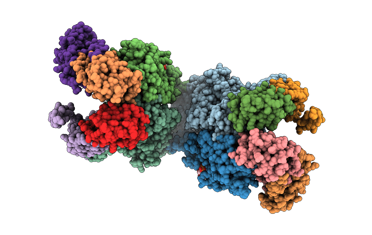
Method: ELECTRON MICROSCOPY Release Date: 2025-03-26 Classification: TOXIN Ligands: CA |
 |
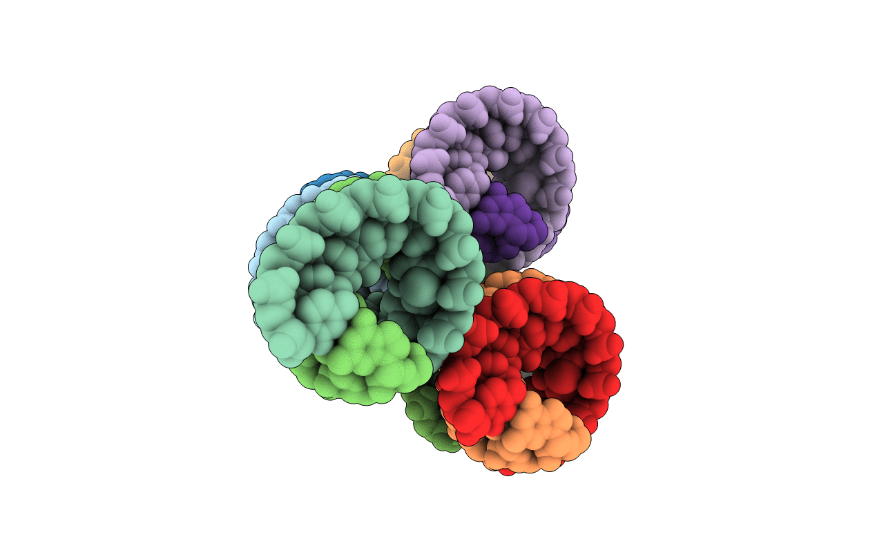
Organism: Synthetic construct
Method: X-RAY DIFFRACTION Resolution:3.10 Å Release Date: 2022-03-16 Classification: RNA |
 |
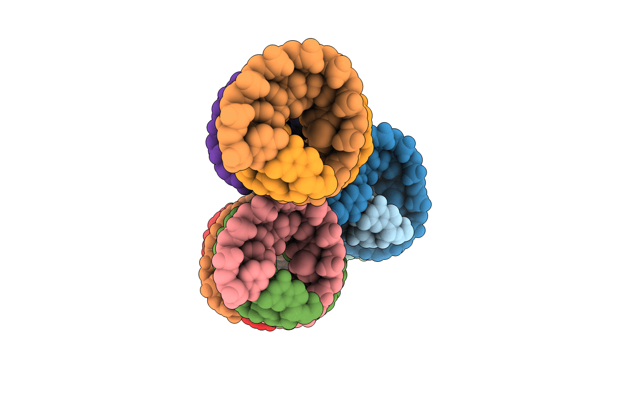
Organism: Synthetic construct
Method: X-RAY DIFFRACTION Resolution:2.79 Å Release Date: 2022-03-16 Classification: RNA Ligands: AG |
 |
Organism: Synthetic construct
Method: X-RAY DIFFRACTION Resolution:3.01 Å Release Date: 2022-03-16 Classification: RNA Ligands: AG |
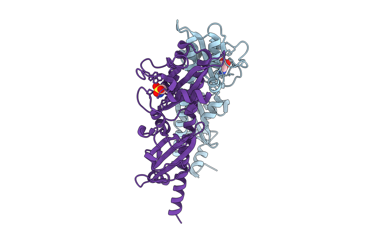 |
Organism: Vibrio cholerae
Method: X-RAY DIFFRACTION Resolution:1.95 Å Release Date: 2016-06-01 Classification: SIGNALING PROTEIN Ligands: TAU |
 |
Organism: Salmonella typhimurium (strain lt2 / sgsc1412 / atcc 700720), Salmonella typhimurium
Method: X-RAY DIFFRACTION Resolution:3.00 Å Release Date: 2016-03-23 Classification: HYDROLASE/MOTOR PROTEIN Ligands: ADP |
 |
Organism: Vibrio cholerae
Method: X-RAY DIFFRACTION Resolution:1.80 Å Release Date: 2016-03-02 Classification: SIGNALING PROTEIN Ligands: SER |
 |
Organism: Plasmid r64
Method: X-RAY DIFFRACTION Resolution:1.50 Å Release Date: 2015-06-10 Classification: UNKNOWN FUNCTION Ligands: SO4 |
 |
Organism: Legionella pneumophila
Method: X-RAY DIFFRACTION Resolution:2.20 Å Release Date: 2015-06-10 Classification: UNKNOWN FUNCTION Ligands: MRD, MPD |
 |
Organism: Legionella pneumophila
Method: X-RAY DIFFRACTION Resolution:3.50 Å Release Date: 2015-06-10 Classification: UNKNOWN FUNCTION |
 |
Crystal Structure Of Mouse Alpha-Tocopherol Transfer Protein In Complex With Alpha-Tocopherol And Phosphatidylinositol-(3,4)-Bisphosphate
Organism: Mus musculus
Method: X-RAY DIFFRACTION Resolution:2.61 Å Release Date: 2013-05-01 Classification: TRANSPORT PROTEIN Ligands: VIV, 3PT |
 |
Crystal Structure Of Mouse Alpha-Tocopherol Transfer Protein In Complex With Alpha-Tocopherol And Phosphatidylinositol-(4,5)-Bisphosphate
Organism: Mus musculus
Method: X-RAY DIFFRACTION Resolution:2.05 Å Release Date: 2013-05-01 Classification: TRANSPORT PROTEIN Ligands: VIV, 4PT, PBU, PO4 |
 |
Organism: Homo sapiens
Method: X-RAY DIFFRACTION Resolution:1.75 Å Release Date: 2012-01-25 Classification: PROTEIN TRANSPORT |
 |
Crystal Structure Of The Ph Domain Of Evectin-2 From Human Complexed With O-Phospho-L-Serine
Organism: Homo sapiens
Method: X-RAY DIFFRACTION Resolution:1.00 Å Release Date: 2011-05-25 Classification: PROTEIN TRANSPORT Ligands: SEP, EDO |
 |
 |
Organism: Pyrococcus horikoshii
Method: X-RAY DIFFRACTION Resolution:1.45 Å Release Date: 2004-11-23 Classification: LIGASE Ligands: ZN |