
Search Count: 27
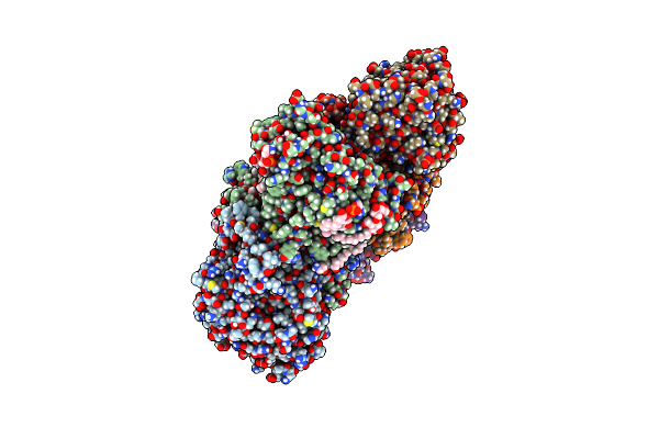 |
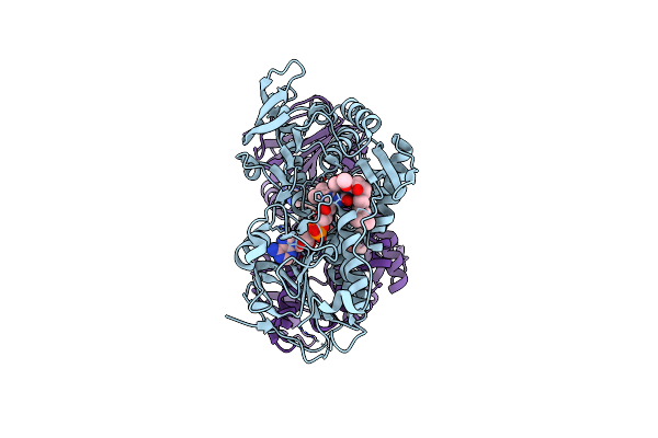
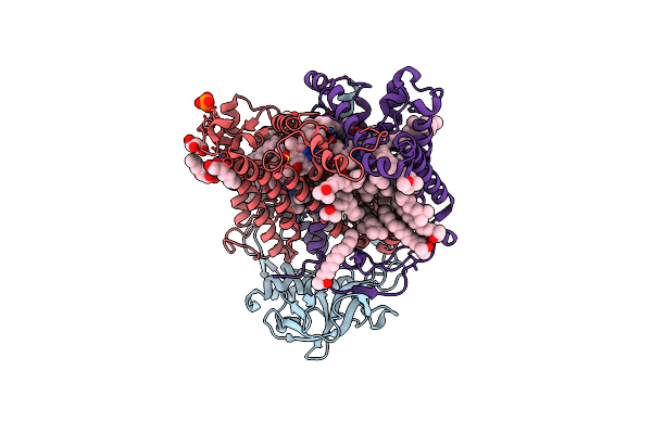
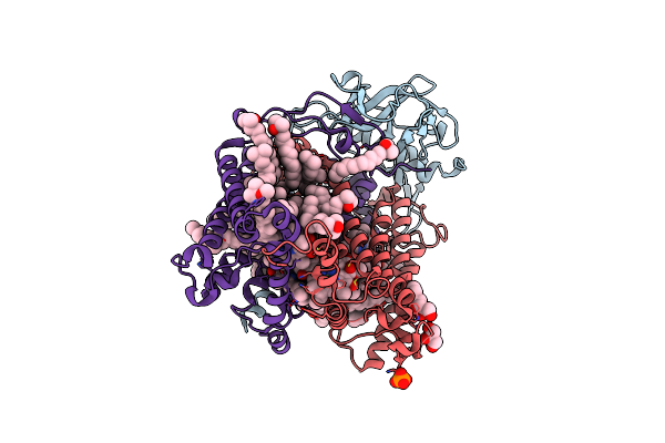
Organism: Vibrio cholerae
Method: ELECTRON MICROSCOPY Release Date: 2023-09-20 Classification: MEMBRANE PROTEIN Ligands: FMN, RBF, LMT, 3PE, UQ2, NA, FES, FAD, NAI |
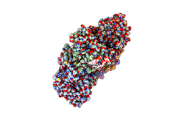 |
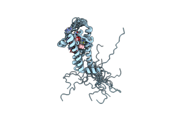
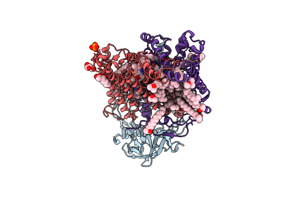
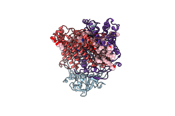
Organism: Vibrio cholerae
Method: ELECTRON MICROSCOPY Release Date: 2023-06-14 Classification: MEMBRANE PROTEIN Ligands: RBF, 3PE, LMT, UQ2, FMN, NA, FES, FAD |
 |
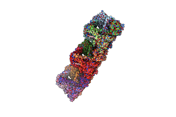
Lipidic Cubic Phase Serial Femtosecond Crystallography Structure Of A Photosynthetic Reaction Centre
Organism: Blastochloris viridis
Method: X-RAY DIFFRACTION Resolution:2.25 Å Release Date: 2022-06-22 Classification: MEMBRANE PROTEIN Ligands: HEC, DGA, LDA, SO4, BCB, BPB, HTO, UQ2, FE2, MQ9, NS5, OLC |
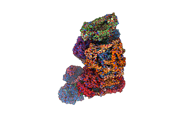 |
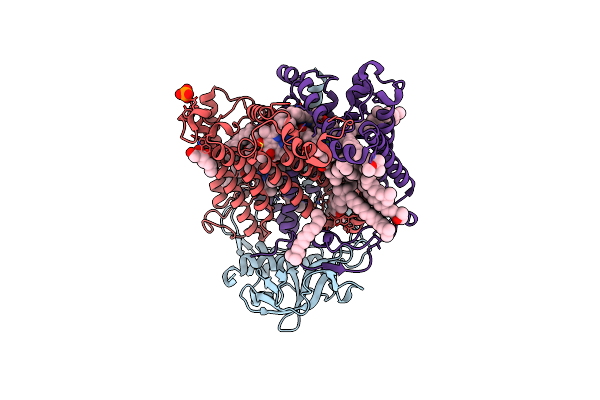
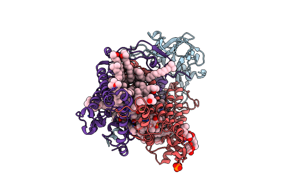
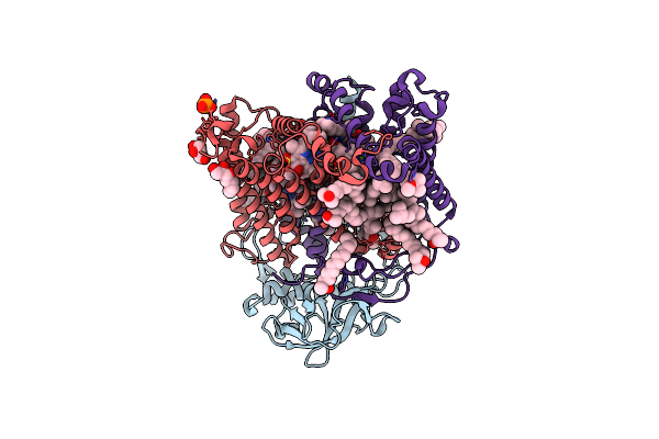
Organism: Bos taurus
Method: ELECTRON MICROSCOPY Release Date: 2022-05-18 Classification: OXIDOREDUCTASE Ligands: PC1, 3PE, FES, CDL, FMN, SF4, CU, HEA, MG, ZN, NAP, HEM, HEC, UQ2 |
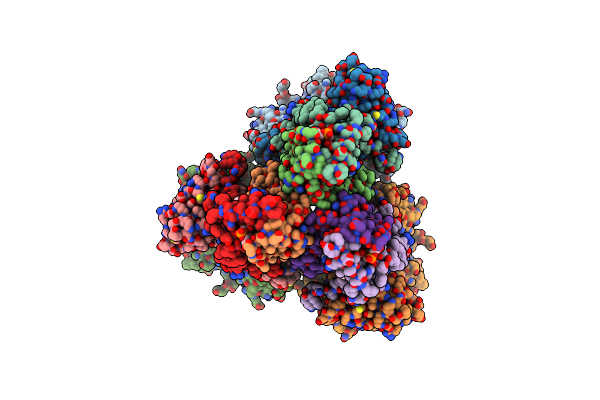 |
Organism: Escherichia coli
Method: ELECTRON MICROSCOPY Release Date: 2021-01-20 Classification: ELECTRON TRANSPORT/OXIDOREDUCTASE Ligands: FAD, NA, FES, SF4, F3S, 3PE, HEM, UQ2 |
 |
Structure Of The Ndi1 Protein From Saccharomyces Cerevisiae In Complex With Quinone
Organism: Saccharomyces cerevisiae
Method: X-RAY DIFFRACTION Resolution:3.00 Å Release Date: 2012-09-05 Classification: OXIDOREDUCTASE Ligands: FAD, UQ2 |
 |
Organism: Escherichia coli
Method: SOLUTION NMR Release Date: 2008-10-07 Classification: MEMBRANE PROTEIN, OXIDOREDUCTASE Ligands: UQ2 |
 |
Crystal Structure Of Rhodobacter Sphaeroides Double Mutant With Stigmatellin And Uq2
Organism: Rhodobacter sphaeroides
Method: X-RAY DIFFRACTION Resolution:2.40 Å Release Date: 2007-12-25 Classification: OXIDOREDUCTASE Ligands: SR, HEM, SMA, LOP, UQ2, BGL, FES, CL, NA |
 |
X-Ray High Resolution Structure Of The Photosynthetic Reaction Center From Rb. Sphaeroides At Ph 6.5 In The Charge-Separated State
Organism: Rhodobacter sphaeroides
Method: X-RAY DIFFRACTION Resolution:2.90 Å Release Date: 2007-07-03 Classification: PHOTOSYNTHESIS Ligands: GOL, LDA, BCL, BPH, UQ2, PO4, FE, U10, SPO |
 |
X-Ray High Resolution Structure Of The Photosynthetic Reaction Center From Rb. Sphaeroides At Ph 6.5 In The Charge-Separated State 2Nd Dataset
Organism: Rhodobacter sphaeroides
Method: X-RAY DIFFRACTION Resolution:2.50 Å Release Date: 2007-07-03 Classification: PHOTOSYNTHESIS Ligands: GOL, BCL, BPH, UQ2, PO4, HTO, LDA, FE, U10, SPO |
 |
X-Ray High Resolution Structure Of The Photosynthetic Reaction Center From Rb. Sphaeroides At Ph 6.5 In The Neutral State, 2Nd Dataset
Organism: Rhodobacter sphaeroides
Method: X-RAY DIFFRACTION Resolution:2.04 Å Release Date: 2007-07-03 Classification: PHOTOSYNTHESIS Ligands: GOL, BCL, LDA, BPH, UQ2, PO4, HTO, FE, U10, SPO, CDL |
 |
X-Ray High Resolution Structure Of The Photosynthetic Reaction Center From Rb. Sphaeroides At Ph 6.5 In The Charge-Separated State, 3Rd Dataset
Organism: Rhodobacter sphaeroides
Method: X-RAY DIFFRACTION Resolution:2.13 Å Release Date: 2007-07-03 Classification: PHOTOSYNTHESIS Ligands: GOL, BCL, LDA, BPH, UQ2, PO4, HTO, FE, U10, SPO |
 |
X-Ray High Resolution Structure Of The Photosynthetic Reaction Center From Rb. Sphaeroides At Ph 6.5 In The Neutral State
Organism: Rhodobacter sphaeroides
Method: X-RAY DIFFRACTION Resolution:2.05 Å Release Date: 2007-07-03 Classification: PHOTOSYNTHESIS Ligands: GOL, BCL, LDA, BPH, UQ2, PO4, HTO, FE, U10, SPO, CDL |
 |
X-Ray High Resolution Structure Of The Photosynthetic Reaction Center From Rb. Sphaeroides At Ph 9 In The Neutral State
Organism: Rhodobacter sphaeroides
Method: X-RAY DIFFRACTION Resolution:2.50 Å Release Date: 2007-07-03 Classification: PHOTOSYNTHESIS Ligands: GOL, BCL, LDA, BPH, UQ2, PO4, HTO, FE, U10, SPO, CDL |
 |
X-Ray High Resolution Structure Of The Photosynthetic Reaction Center From Rb. Sphaeroides At Ph 9 In The Charge-Separated State, 2Nd Dataset
Organism: Rhodobacter sphaeroides
Method: X-RAY DIFFRACTION Resolution:2.51 Å Release Date: 2007-07-03 Classification: PHOTOSYNTHESIS Ligands: GOL, BCL, LDA, BPH, UQ2, PO4, HTO, FE, U10, SPO, CDL |
 |
X-Ray High Resolution Structure Of The Photosynthetic Reaction Center From Rb. Sphaeroides At Ph 9 In The Charge-Separated State
Organism: Rhodobacter sphaeroides
Method: X-RAY DIFFRACTION Resolution:2.21 Å Release Date: 2007-07-03 Classification: PHOTOSYNTHESIS Ligands: GOL, BCL, LDA, BPH, UQ2, PO4, HTO, FE, U10, SPO, CDL |
 |
X-Ray High Resolution Structure Of The Photosynthetic Reaction Center From Rb. Sphaeroides At Ph 10 In The Neutral State
Organism: Rhodobacter sphaeroides
Method: X-RAY DIFFRACTION Resolution:2.25 Å Release Date: 2007-07-03 Classification: PHOTOSYNTHESIS Ligands: GOL, BCL, LDA, BPH, UQ2, PO4, HTO, FE, U10, SPO, CDL |
 |
X-Ray High Resolution Structure Of The Photosynthetic Reaction Center From Rb. Sphaeroides At Ph 10 In The Charge-Separated State
Organism: Rhodobacter sphaeroides
Method: X-RAY DIFFRACTION Resolution:2.31 Å Release Date: 2007-07-03 Classification: PHOTOSYNTHESIS Ligands: GOL, BCL, LDA, BPH, UQ2, PO4, HTO, FE, U10, SPO, CDL |
 |
X-Ray High Resolution Structure Of The Photosynthetic Reaction Center From Rb. Sphaeroides At Ph 10 In The Neutral State, 2Nd Dataset
Organism: Rhodobacter sphaeroides
Method: X-RAY DIFFRACTION Resolution:2.88 Å Release Date: 2007-07-03 Classification: PHOTOSYNTHESIS Ligands: GOL, BCL, BPH, UQ2, HTO, LDA, FE, U10, SPO |
 |
X-Ray High Resolution Structure Of The Photosynthetic Reaction Center From Rb. Sphaeroides At Ph 10 In The Charge-Separated State, 2Nd Dataset
Organism: Rhodobacter sphaeroides
Method: X-RAY DIFFRACTION Resolution:2.70 Å Release Date: 2007-07-03 Classification: PHOTOSYNTHESIS Ligands: GOL, BCL, LDA, BPH, UQ2, PO4, HTO, FE, U10, SPO |