
Search Count: 47
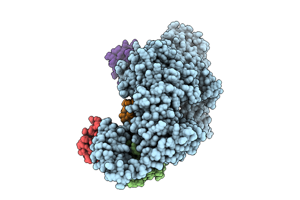 |
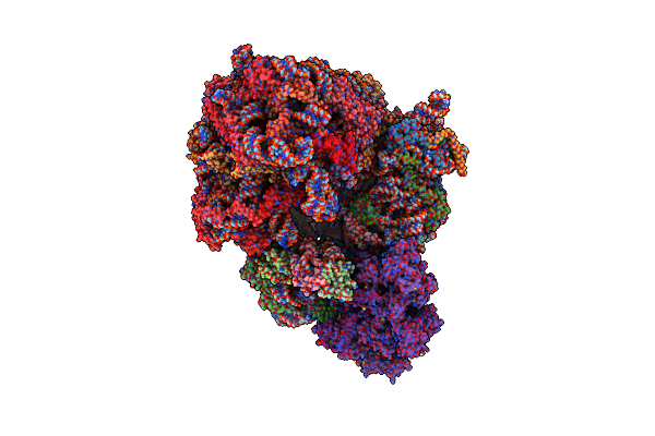
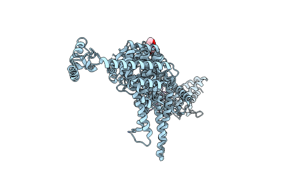
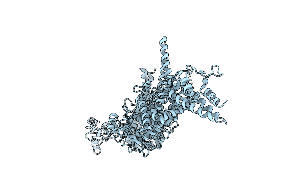
Cryo-Em Structure Of Ufd2/Ubc4-Ub In Complex With K29-Linked Diub (Monomeric Conformation)
Organism: Saccharomyces cerevisiae (strain atcc 204508 / s288c), Homo sapiens
Method: ELECTRON MICROSCOPY Release Date: 2025-07-30 Classification: LIGASE |
 |
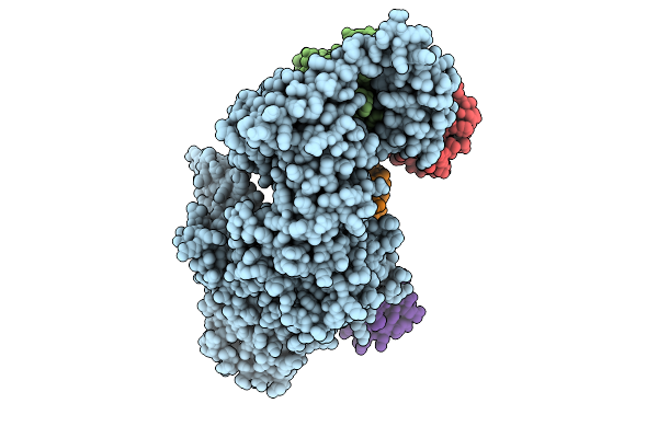
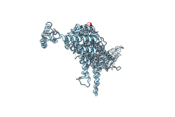
Cryo-Em Structure Of Ufd2/Ubc4-Ub In Complex With K29-Linked Triub (Dimeric Conformation)
Organism: Homo sapiens, Saccharomyces cerevisiae (strain atcc 204508 / s288c)
Method: ELECTRON MICROSCOPY Release Date: 2025-07-30 Classification: LIGASE |
 |
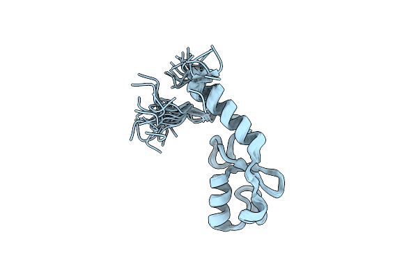
Cryo-Em Structure Of Ufd2/Ubc4-Ub Complex With K29Triub(Monomeric Conformation)
Organism: Saccharomyces cerevisiae (strain atcc 204508 / s288c), Homo sapiens
Method: ELECTRON MICROSCOPY Release Date: 2025-07-30 Classification: LIGASE |
 |
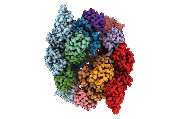
Organism: Saccharomyces cerevisiae
Method: ELECTRON MICROSCOPY Release Date: 2023-02-22 Classification: RIBOSOME Ligands: MG, ZN |
 |
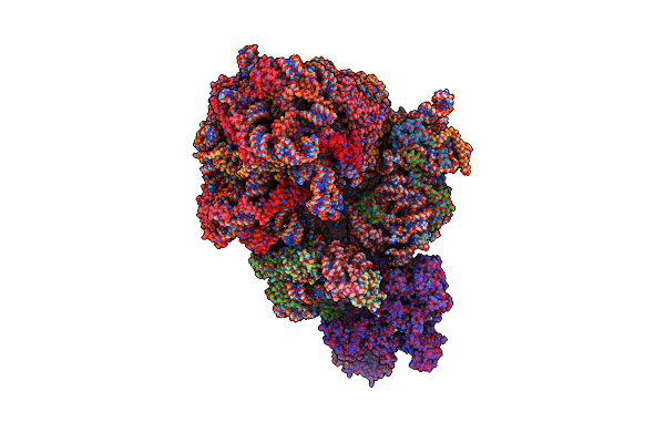
Structure Of The Rqt-Bound 80S Ribosome From S. Cerevisiae (C2) - Composite Map
Organism: Saccharomyces cerevisiae
Method: ELECTRON MICROSCOPY Release Date: 2023-02-22 Classification: RIBOSOME Ligands: MG, ZN |
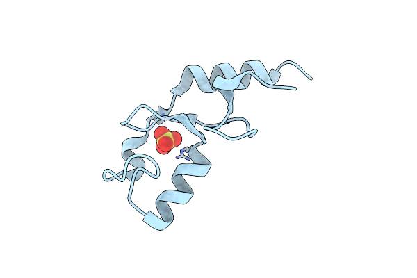 |
Organism: Homo sapiens
Method: X-RAY DIFFRACTION Resolution:1.48 Å Release Date: 2017-11-01 Classification: LIGASE Ligands: SO4 |
 |
Organism: Saccharomyces cerevisiae s288c
Method: SOLUTION NMR Release Date: 2016-03-23 Classification: UBIQUITIN-BINDING PROTEIN |
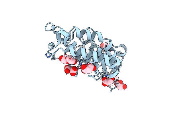 |
Structure Of Ube2Z Provides Functional Insight Into Specificity In The Fat10 Conjugation Machinery
Organism: Homo sapiens
Method: X-RAY DIFFRACTION Resolution:2.10 Å Release Date: 2015-11-18 Classification: LIGASE Ligands: MLI, PEG |
 |
Crystal Structure Of Ehubc5, A Ubiquitin Conjugating Enzyme From Entamoeba Histolytica
Organism: Entamoeba histolytica
Method: X-RAY DIFFRACTION Resolution:1.60 Å Release Date: 2012-12-12 Classification: LIGASE Ligands: CO |
 |
Organism: Kluyveromyces marxianus
Method: X-RAY DIFFRACTION Resolution:3.20 Å Release Date: 2012-11-14 Classification: LIGASE |
 |
Organism: Homo sapiens
Method: X-RAY DIFFRACTION Resolution:2.60 Å Release Date: 2010-05-05 Classification: LIGASE |
 |
Crystal Structure Of Ufd2 In Complex With The Ubiquitin-Like (Ubl) Domain Of Rad23
Organism: Saccharomyces cerevisiae
Method: X-RAY DIFFRACTION Resolution:2.40 Å Release Date: 2010-04-28 Classification: LIGASE/PROTEIN BINDING Ligands: 1PE, K |
 |
Crystal Structure Of Ufd2 In Complex With The Ubiquitin-Like (Ubl) Domain Of Dsk2
Organism: Saccharomyces cerevisiae
Method: X-RAY DIFFRACTION Resolution:2.40 Å Release Date: 2010-04-28 Classification: LIGASE/PROTEIN BINDING Ligands: 1PE, K |
 |
 |
Organism: Homo sapiens
Method: SOLUTION NMR Release Date: 2009-12-29 Classification: PROTEIN BINDING |
 |
Organism: Saccharomyces cerevisiae
Method: X-RAY DIFFRACTION Resolution:2.56 Å Release Date: 2007-09-18 Classification: LIGASE Ligands: K |
 |
Organism: Saccharomyces cerevisiae
Method: X-RAY DIFFRACTION Resolution:2.65 Å Release Date: 2007-09-18 Classification: LIGASE |
 |
Organism: Homo sapiens
Method: SOLUTION NMR Release Date: 2004-11-28 Classification: PROTEIN BINDING |
 |
 |
E2-C, An Ubiquitin Conjugating Enzyme Required For The Destruction Of Mitotic Cyclins
Organism: Spisula solidissima
Method: X-RAY DIFFRACTION Resolution:2.00 Å Release Date: 1999-01-27 Classification: UBIQUITIN CONJUGATION |