
Search Count: 19
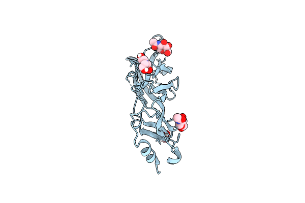 |
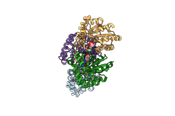
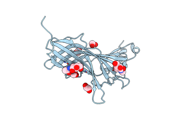
Crystal Structure Of The Beta3 Subunit Extracellular Domain Of Nicotinic Acetylcholine Receptor
Organism: Homo sapiens
Method: X-RAY DIFFRACTION Resolution:2.40 Å Release Date: 2022-08-10 Classification: MEMBRANE PROTEIN Ligands: NAG, BCN, EDO |
 |
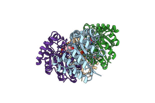
Organism: Bacillus anthracis
Method: X-RAY DIFFRACTION Resolution:2.59 Å Release Date: 2019-04-10 Classification: HYDROLASE Ligands: ZN, ACT, EDO, SO4 |
 |
Crystal Structure Of The Polysaccharide Deacetylase Bc1974 From Bacillus Cereus
Organism: Bacillus cereus
Method: X-RAY DIFFRACTION Resolution:1.80 Å Release Date: 2018-02-21 Classification: HYDROLASE Ligands: ZN, ACT, EDO |
 |
Crystal Structure Of The Polysaccharide Deacetylase Bc1974 From Bacillus Cereus In Complex With N-Hydroxynaphthalene-1-Carboxamide
Organism: Bacillus cereus
Method: X-RAY DIFFRACTION Resolution:1.45 Å Release Date: 2018-02-21 Classification: HYDROLASE Ligands: 8GK, ZN, EDO, NA, PGE |
 |
Crystal Structure Of The Polysaccharide Deacetylase Bc1974 From Bacillus Cereus In Complex With (E)-N-Hydroxy-3-(Naphthalen-1-Yl)Prop-2-Enamide
Organism: Bacillus cereus
Method: X-RAY DIFFRACTION Resolution:2.80 Å Release Date: 2018-02-21 Classification: HYDROLASE Ligands: ACT, ZN, EDO, 8SQ, PGE |
 |
Crystal Structure Of The Polysaccharide Deacetylase Bc1974 From Bacillus Cereus In Complex With (2S)-2,6-Diamino-N-Hydroxyhexanamide
Organism: Bacillus cereus atcc 14579, Bacillus cereus
Method: X-RAY DIFFRACTION Resolution:2.44 Å Release Date: 2018-02-21 Classification: HYDROLASE Ligands: 8SZ, ZN, CIT, EDO, PGE |
 |
Crystal Structure Of The Polysaccharide Deacetylase Bc1974 From Bacillus Cereus In Complex With (2S)-2-Amino-5-(Diaminomethylideneamino)-N-Hydroxypentanamide
Organism: Bacillus cereus
Method: X-RAY DIFFRACTION Resolution:2.45 Å Release Date: 2018-02-21 Classification: HYDROLASE Ligands: ACT, ZN, AHL, CIT |
 |
Crystal Structure Of The Polysaccharide Deacetylase Bc1974 From Bacillus Cereus In Complex With Acetazolamide
Organism: Bacillus cereus
Method: X-RAY DIFFRACTION Resolution:3.06 Å Release Date: 2018-02-21 Classification: HYDROLASE Ligands: ZN, ACT, AZM, PGE |
 |
Crystal Structure Of The Polysaccharide Deacetylase Bc1974 From Bacillus Cereus In Complex With Thiametg
Organism: Bacillus cereus
Method: X-RAY DIFFRACTION Resolution:2.73 Å Release Date: 2018-02-21 Classification: HYDROLASE Ligands: ZN, ACT, NHT, PGE, EDO, CIT |
 |
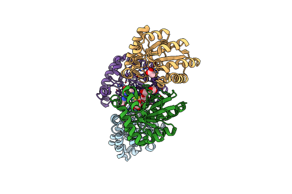
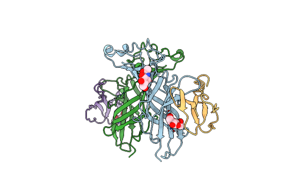
Crystal Structure Of The Extracellular Domain Of Alpha2 Nicotinic Acetylcholine Receptor In Pentameric Assembly
Organism: Homo sapiens
Method: X-RAY DIFFRACTION Resolution:3.20 Å Release Date: 2016-08-10 Classification: ACETYLCHOLINE-BINDING PROTEIN Ligands: EPJ |
 |
Crystal Structure Of The Putative Polysaccharide Deacetylase Ba0330 From Bacillus Anthracis
Organism: Bacillus anthracis
Method: X-RAY DIFFRACTION Resolution:1.48 Å Release Date: 2015-04-08 Classification: HYDROLASE Ligands: ACT, ZN, EDO |
 |
Crystal Structure Of The Extracellular Domain Of The Human Alpha9 Nicotinic Acetylcholine Receptor
Organism: Homo sapiens
Method: X-RAY DIFFRACTION Resolution:1.80 Å Release Date: 2014-10-01 Classification: SIGNALING PROTEIN Ligands: NAG, EDO |
 |
Crystal Structure Of The Extracellular Domain Of The Human Alpha9 Nicotinic Acetylcholine Receptor In Complex With Methyllycaconitine
Organism: Homo sapiens
Method: X-RAY DIFFRACTION Resolution:1.71 Å Release Date: 2014-10-01 Classification: TRANSPORT PROTEIN Ligands: MLK, EPE, EDO, NA, NAG |
 |
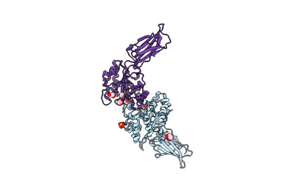
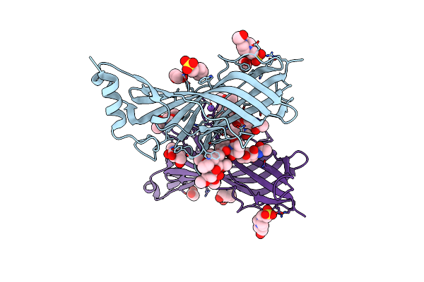
Crystal Structure Of The Complex Of The Extracellular Domain Of Human Alpha9 Nachr With Alpha-Bungarotoxin.
Organism: Homo sapiens, Bungarus multicinctus
Method: X-RAY DIFFRACTION Resolution:2.70 Å Release Date: 2014-10-01 Classification: TOXIN-BINDING PROTEIN/TOXIN Ligands: NAG |
 |
Crystal Structure Of Fab198, An Efficient Protector Of Acetylcholine Receptor Against Myasthenogenic Antibodies
Organism: Rattus norvegicus
Method: X-RAY DIFFRACTION Resolution:2.80 Å Release Date: 2001-09-26 Classification: IMMUNE SYSTEM |
 |
Complex Between Fv Antibody Fragment And An Analogue Of The Main Immunogenic Region Of The Acetylcholine Receptor
Organism: Rattus norvegicus
Method: SOLUTION NMR Release Date: 2000-06-15 Classification: IMMUNE SYSTEM |
 |
The Crystal Structure Of The Fab Fragment Of A Rat Monoclonal Antibody Against The Main Immunogenic Region Of The Human Muscle Acetylcholine Receptor
Organism: Rattus norvegicus
Method: X-RAY DIFFRACTION Resolution:2.40 Å Release Date: 1999-12-03 Classification: IMMUNE SYSTEM |
 |
Molecular Dynamics Simulation From 2D-Nmr Data Of The Free Achr Mir Decapeptide And The Antibody-Bound [A76]Mir Analogue
Organism: Torpedo californica
Method: SOLUTION NMR Release Date: 1996-03-08 Classification: TRANSMEMBRANE PROTEIN |
 |
Torpedo Californica Achr Receptor [Ala76] Analogue Complexed With The Anti-Acetylcholine Mab6 Monoclonal Antibody
Organism: Torpedo californica
Method: SOLUTION NMR Release Date: 1996-03-08 Classification: TRANSMEMBRANE PROTEIN |