
Search Count: 12
 |
Organism: Panicum virgatum
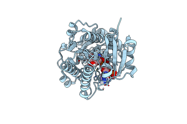
Method: X-RAY DIFFRACTION Resolution:2.34 Å Release Date: 2023-09-20 Classification: OXIDOREDUCTASE Ligands: NAP |
 |
Organism: Panicum virgatum
Method: X-RAY DIFFRACTION Resolution:2.55 Å Release Date: 2023-09-20 Classification: OXIDOREDUCTASE Ligands: NAP, DQH |
 |
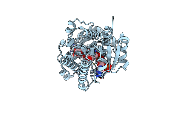
Organism: Sorghum bicolor
Method: X-RAY DIFFRACTION Resolution:2.20 Å Release Date: 2023-09-20 Classification: OXIDOREDUCTASE Ligands: NAP |
 |
Organism: Sorghum bicolor
Method: X-RAY DIFFRACTION Resolution:2.12 Å Release Date: 2023-09-20 Classification: OXIDOREDUCTASE Ligands: NAR, NAP, SO4 |
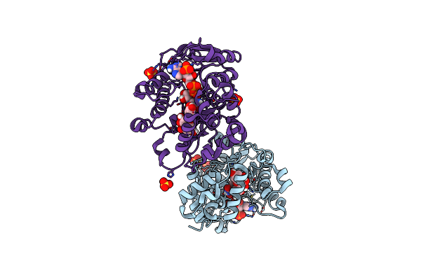 |
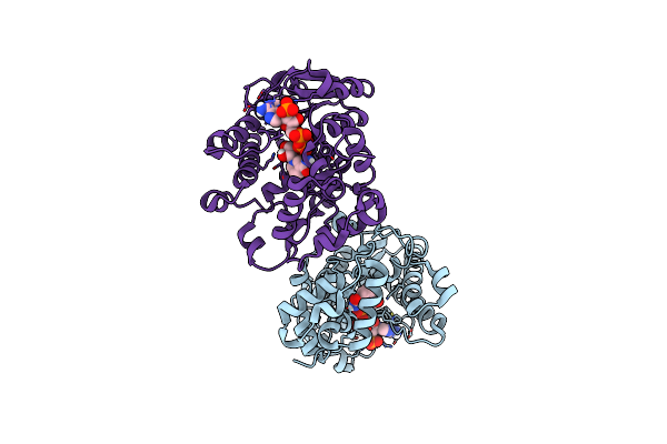
Flavanone 4-Reductase From Sorghum Bicolor-Nadp(H) And Dihydroquercetin Complex
Organism: Sorghum bicolor
Method: X-RAY DIFFRACTION Resolution:2.21 Å Release Date: 2023-09-20 Classification: OXIDOREDUCTASE Ligands: DQH, NAP, SO4 |
 |
Organism: Sorghum bicolor
Method: X-RAY DIFFRACTION Resolution:2.02 Å Release Date: 2023-09-20 Classification: OXIDOREDUCTASE Ligands: NAR, SO4 |
 |
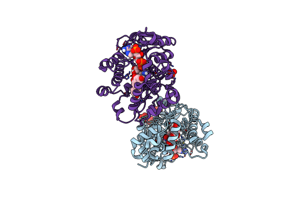
Hypothetical Anthocyanidin Reductase From Sorghum Bicolor-Nadp(H) And Naringenin Complex
Organism: Sorghum bicolor
Method: X-RAY DIFFRACTION Resolution:1.97 Å Release Date: 2023-09-20 Classification: OXIDOREDUCTASE Ligands: NAR, NAP |
 |
Organism: Sorghum bicolor
Method: X-RAY DIFFRACTION Resolution:1.70 Å Release Date: 2023-09-20 Classification: OXIDOREDUCTASE Ligands: NAP |
 |
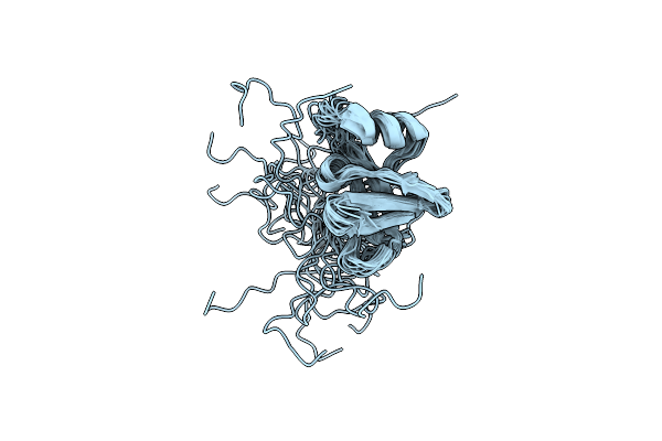
Organism: Homo sapiens
Method: SOLUTION NMR Release Date: 2013-01-23 Classification: PROTEIN TRANSPORT Ligands: ZN |
 |
Crystal Structure Of Ubiquitin-Conjugating Enzyme E2-25Kda (Huntington Interacting Protein 2) M172A Mutant Crystallized At Ph 8.5
Organism: Homo sapiens
Method: X-RAY DIFFRACTION Resolution:2.23 Å Release Date: 2008-11-25 Classification: LIGASE Ligands: PG4, BME, TRS, CA |
 |
Crystal Structure Of Ubiquitin-Conjugating Enzyme E2-25Kda (Huntington Interacting Protein 2) M172A Mutant
Organism: Homo sapiens
Method: X-RAY DIFFRACTION Resolution:1.86 Å Release Date: 2008-08-26 Classification: LIGASE Ligands: CA |
 |
Organism: Corynebacterium diphtheriae
Method: SOLUTION NMR Release Date: 1998-10-21 Classification: GENE REGULATION |