
Search Count: 27
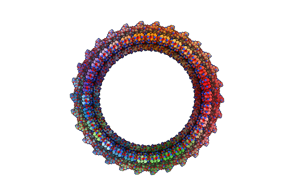 |
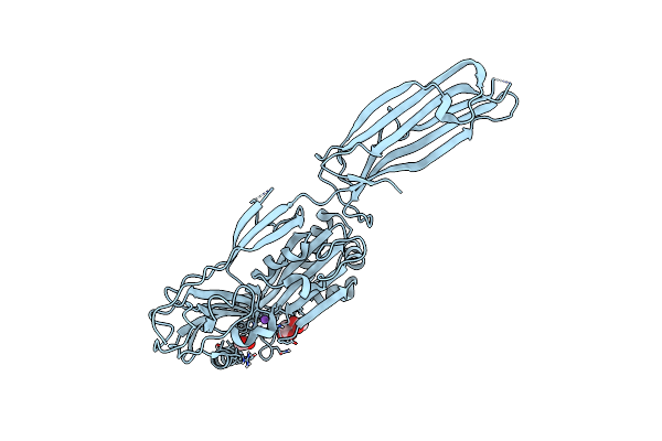
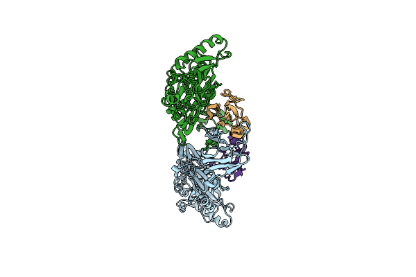
Organism: Elizabethkingia anophelis ag1
Method: ELECTRON MICROSCOPY Release Date: 2025-04-09 Classification: TOXIN Ligands: CA |
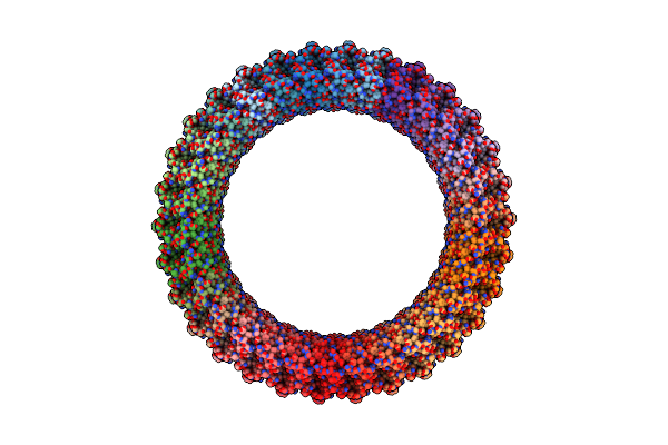 |
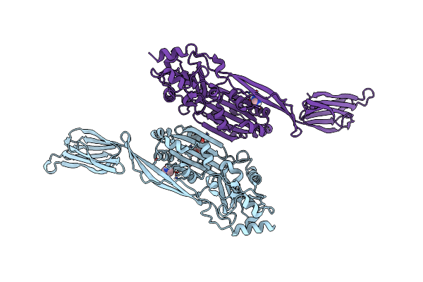
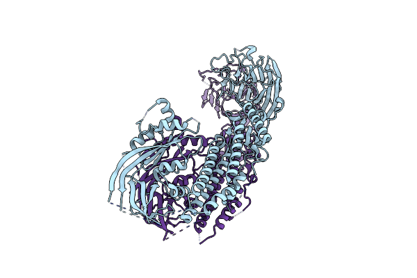
Organism: Elizabethkingia anophelis ag1
Method: ELECTRON MICROSCOPY Release Date: 2025-04-09 Classification: TOXIN Ligands: CA |
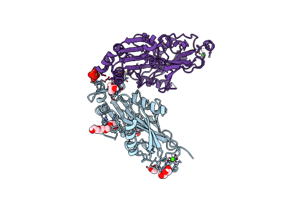 |
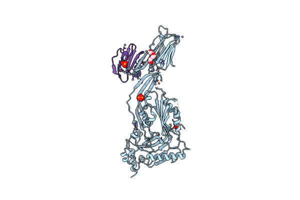
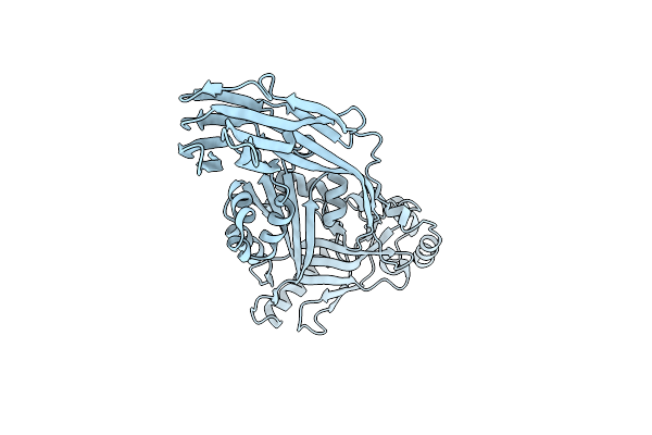
Organism: Elizabethkingia anophelis ag1
Method: X-RAY DIFFRACTION Resolution:1.85 Å Release Date: 2024-02-07 Classification: TOXIN Ligands: SO4, GOL, EDO, PG4, CA, NA |
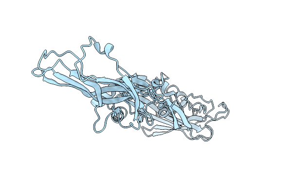 |
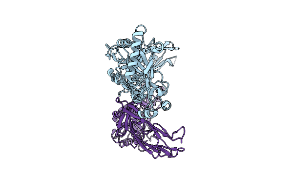
Organism: Elizabethkingia anophelis ag1
Method: X-RAY DIFFRACTION Resolution:2.49 Å Release Date: 2024-02-07 Classification: TOXIN |
 |
Organism: Elizabethkingia anophelis
Method: X-RAY DIFFRACTION Resolution:2.10 Å Release Date: 2021-04-21 Classification: TOXIN |
 |
Organism: Desulfobulbus propionicus (strain atcc 33891 / dsm 2032 / 1pr3)
Method: X-RAY DIFFRACTION Resolution:2.30 Å Release Date: 2019-12-04 Classification: TOXIN Ligands: PG4, IMD |
 |
Organism: Streptococcus intermedius, Homo sapiens
Method: X-RAY DIFFRACTION Resolution:2.70 Å Release Date: 2016-08-24 Classification: TOXIN Ligands: PGE, SO4, ZN, CU |
 |
Organism: Streptococcus intermedius
Method: X-RAY DIFFRACTION Resolution:2.89 Å Release Date: 2016-08-24 Classification: TOXIN |
 |
Organism: Gardnerella vaginalis, Homo sapiens
Method: X-RAY DIFFRACTION Resolution:2.40 Å Release Date: 2016-08-24 Classification: toxin/toxin receptor |
 |
Organism: Streptococcus pneumoniae
Method: X-RAY DIFFRACTION Resolution:2.90 Å Release Date: 2016-03-09 Classification: Sugar binding protein, toxin Ligands: EDO, PEG, AUC |
 |
Organism: Synanceia horrida
Method: X-RAY DIFFRACTION Resolution:3.10 Å Release Date: 2015-12-02 Classification: TOXIN |
 |
Structure Of Membrane Binding Protein Pleurotolysin A From Pleurotus Ostreatus
Organism: Pleurotus ostreatus
Method: X-RAY DIFFRACTION Resolution:1.85 Å Release Date: 2015-02-18 Classification: MEMBRANE BINDING PROTEIN Ligands: SO4 |
 |
Structure Of Membrane Binding Protein Pleurotolysin B From Pleurotus Ostreatus
Organism: Pleurotus ostreatus
Method: X-RAY DIFFRACTION Resolution:2.20 Å Release Date: 2015-02-18 Classification: MEMBRANE BINDING PROTEIN Ligands: ACT, GOL, CL |
 |
Crystal Structure Of The Tmh1-Lock Mutant Of The Mature Form Of Pleurotolysin B
Organism: Pleurotus ostreatus
Method: X-RAY DIFFRACTION Resolution:2.15 Å Release Date: 2015-02-18 Classification: TOXIN Ligands: SO4, CL, GOL |
 |
Organism: Pleurotus ostreatus
Method: ELECTRON MICROSCOPY Resolution:11.00 Å Release Date: 2015-02-18 Classification: TRANSPORT PROTEIN |
 |
Membrane Bound Pleurotolysin Prepore (Tmh1 Lock) Trapped With Engineered Disulphide Cross-Link
Organism: Pleurotus ostreatus
Method: ELECTRON MICROSCOPY Resolution:15.00 Å Release Date: 2015-02-18 Classification: TRANSPORT PROTEIN |
 |
Membrane Bound Pleurotolysin Prepore (Tmh2 Helix Lock) Trapped With Engineered Disulphide Cross-Link
Organism: Pleurotus ostreatus
Method: ELECTRON MICROSCOPY Resolution:17.00 Å Release Date: 2015-02-18 Classification: TRANSPORT PROTEIN |
 |
Membrane Bound Pleurotolysin Prepore (Tmh2 Strand Lock) Trapped With Engineered Disulphide Cross-Link
Organism: Pleurotus ostreatus
Method: ELECTRON MICROSCOPY Resolution:14.00 Å Release Date: 2015-02-18 Classification: TRANSPORT PROTEIN |
 |
Organism: Streptococcus pyogenes
Method: X-RAY DIFFRACTION Resolution:2.10 Å Release Date: 2013-10-30 Classification: TOXIN |
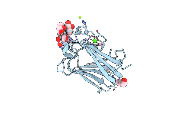 |
His 62 Mutant Of The Lectin Binding Domain Of Lectinolysin Complexed With Lewis Y
Organism: Streptococcus mitis
Method: X-RAY DIFFRACTION Resolution:1.60 Å Release Date: 2012-11-21 Classification: SUGAR BINDING PROTEIN Ligands: MG, GOL, CA |