
Search Count: 27
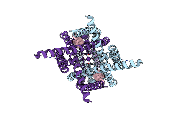 |
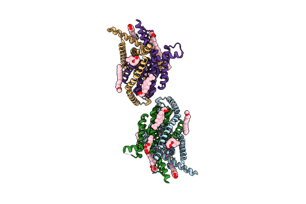
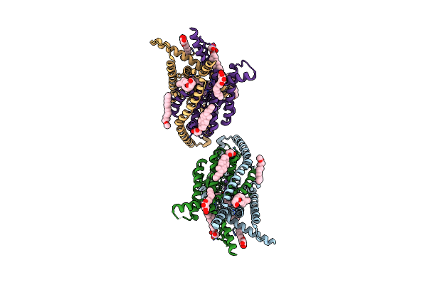
Structure Of The Human Two Pore Domain Potassium Ion Channel Thik-1 (K2P13.1) In A Closed Conformation
Organism: Homo sapiens
Method: ELECTRON MICROSCOPY Release Date: 2025-03-05 Classification: MEMBRANE PROTEIN Ligands: EIC, K |
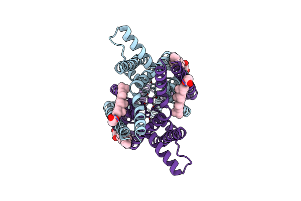 |
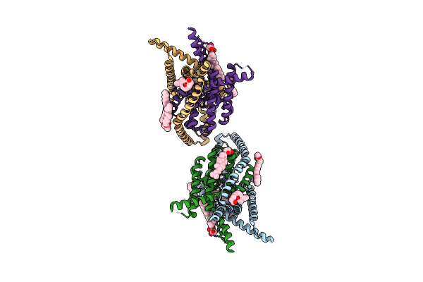
Structure Of The Human Two Pore Domain Potassium Ion Channel Task-3 (K2P9.1)
Organism: Homo sapiens
Method: ELECTRON MICROSCOPY Release Date: 2024-12-18 Classification: MEMBRANE PROTEIN Ligands: Y01, K |
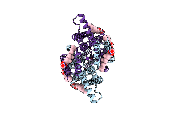 |
Structure Of The Human Two Pore Domain Potassium Ion Channel Task-3 (K2P9.1) G236R Mutant
Organism: Homo sapiens
Method: ELECTRON MICROSCOPY Release Date: 2024-12-18 Classification: MEMBRANE PROTEIN Ligands: Y01, K |
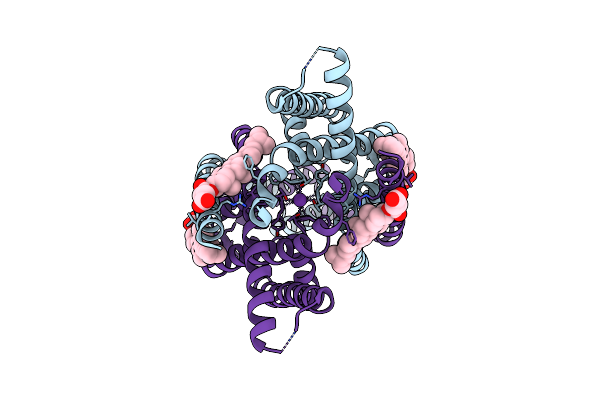 |
Structure Of The Human Two Pore Domain Potassium Ion Channel Task-1 (K2P3.1)
Organism: Homo sapiens
Method: ELECTRON MICROSCOPY Release Date: 2024-12-18 Classification: MEMBRANE PROTEIN Ligands: Y01, K |
 |
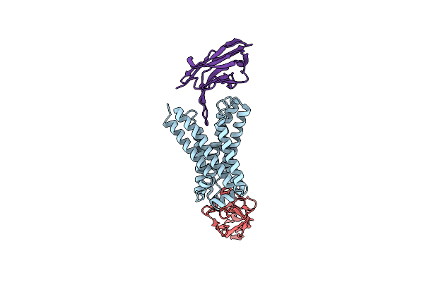
Crystal Structure Of Human Two Pore Domain Potassium Ion Channel Trek-2 (K2P10.1) In Complex With A Nanobody (Nb58)
Organism: Homo sapiens, Lama glama
Method: X-RAY DIFFRACTION Resolution:3.59 Å Release Date: 2024-05-29 Classification: MEMBRANE PROTEIN Ligands: K |
 |
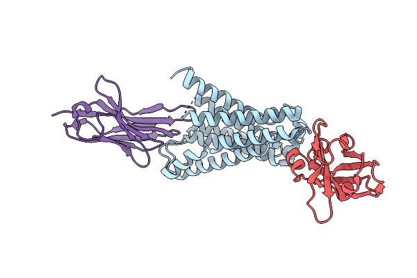
Crystal Structure Of Human Two Pore Domain Potassium Ion Channel Trek-2 (K2P10.1) In Complex With An Inhibitory Nanobody (Nb61)
Organism: Homo sapiens, Lama glama
Method: X-RAY DIFFRACTION Resolution:3.50 Å Release Date: 2024-05-29 Classification: MEMBRANE PROTEIN Ligands: K |
 |
Crystal Structure Of Human Two Pore Domain Potassium Ion Channel Trek-2 (K2P10.1) In Complex With An Activatory Nanobody (Nb67)
Organism: Homo sapiens, Lama glama
Method: X-RAY DIFFRACTION Resolution:2.40 Å Release Date: 2024-05-29 Classification: MEMBRANE PROTEIN Ligands: K, MPD |
 |
Crystal Structure Of Human Two Pore Domain Potassium Ion Channel Trek-2 (K2P10.1) In Complex With An Activatory Nanobody (Nb76)
Organism: Homo sapiens, Lama glama
Method: X-RAY DIFFRACTION Resolution:3.20 Å Release Date: 2024-05-29 Classification: MEMBRANE PROTEIN Ligands: BA, Y01 |
 |
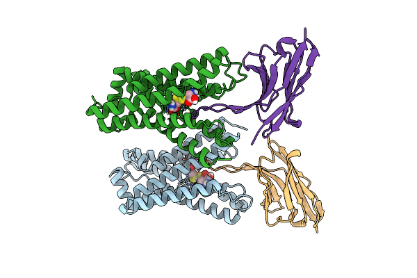
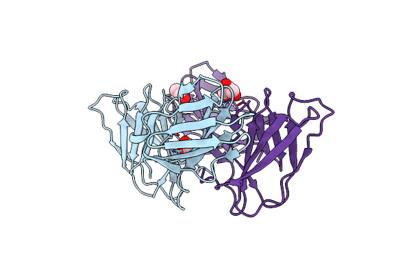
Crystal Structure Of Cystinosin From Arabidopsis Thaliana Bound To Sybody And Nanobody
Organism: Arabidopsis thaliana, Lama glama, Synthetic construct
Method: X-RAY DIFFRACTION Resolution:2.65 Å Release Date: 2022-08-31 Classification: MEMBRANE PROTEIN |
 |
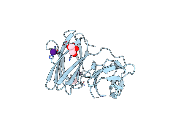
Crystal Structure Of Cystinosin From Arabidopsis Thaliana In Complex With Cystine And Sybody
Organism: Arabidopsis thaliana, Synthetic construct
Method: X-RAY DIFFRACTION Resolution:3.37 Å Release Date: 2022-08-31 Classification: MEMBRANE PROTEIN Ligands: IYY |
 |
Crystal Structure Of Cystinosin From Arabidopsis Thaliana Bound To Two Nanobodies
Organism: Arabidopsis thaliana, Lama glama
Method: X-RAY DIFFRACTION Resolution:2.33 Å Release Date: 2022-08-31 Classification: MEMBRANE PROTEIN |
 |
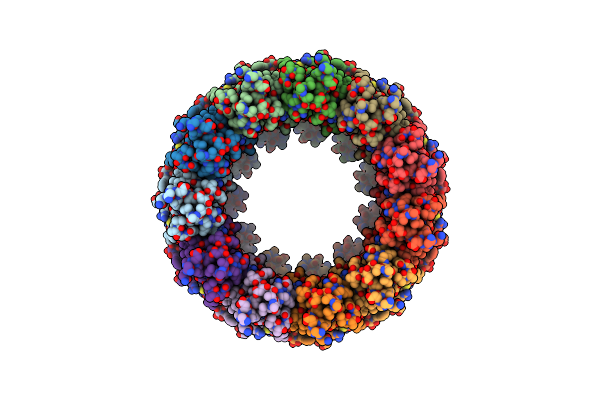
Organism: Homo sapiens
Method: ELECTRON MICROSCOPY Release Date: 2020-01-29 Classification: MEMBRANE PROTEIN |
 |
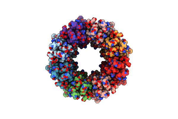
Organism: Homo sapiens
Method: ELECTRON MICROSCOPY Release Date: 2020-01-29 Classification: MEMBRANE PROTEIN |
 |
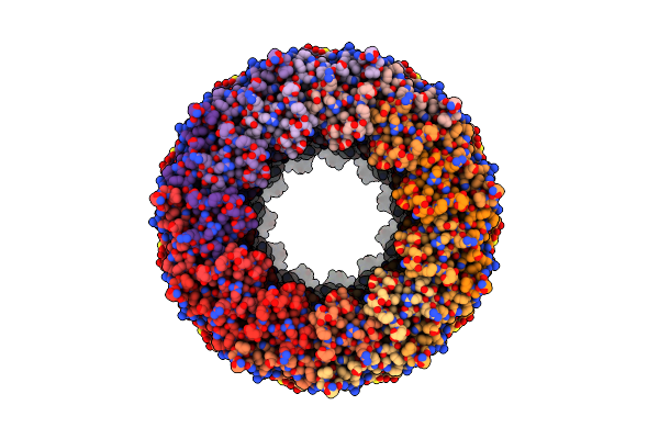
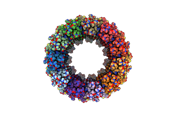
Cryo-Em Structure Of An Undecameric Chicken Calhm1 And Human Calhm2 Chimera
Organism: Aequorea victoria, Gallus gallus, Homo sapiens
Method: ELECTRON MICROSCOPY Release Date: 2020-01-29 Classification: MEMBRANE PROTEIN |
 |
Organism: Aequorea victoria, Gallus gallus
Method: ELECTRON MICROSCOPY Release Date: 2020-01-29 Classification: MEMBRANE PROTEIN |
 |
Crystal Structure Of The Human Two Pore Domain Potassium Ion Channel Task-1 (K2P3.1) In A Closed Conformation
Organism: Homo sapiens
Method: X-RAY DIFFRACTION Resolution:3.00 Å Release Date: 2019-08-07 Classification: MEMBRANE PROTEIN Ligands: K, Y01, DMU, PC1 |
 |
Crystal Structure Of The Human Two Pore Domain Potassium Ion Channel Task-1 (K2P3.1) In A Closed Conformation With A Bound Inhibitor Bay 1000493
Organism: Homo sapiens
Method: X-RAY DIFFRACTION Resolution:2.90 Å Release Date: 2019-08-07 Classification: MEMBRANE PROTEIN Ligands: K, Y01, DMU, PC1, KKQ |
 |
Crystal Structure Of The Human Two Pore Domain Potassium Ion Channel Task-1 (K2P3.1) In A Closed Conformation With A Bound Inhibitor Bay 2341237
Organism: Homo sapiens
Method: X-RAY DIFFRACTION Resolution:3.10 Å Release Date: 2019-08-07 Classification: MEMBRANE PROTEIN Ligands: K, Y01, KKZ, PC1 |
 |
Organism: Mus musculus
Method: X-RAY DIFFRACTION Resolution:2.10 Å Release Date: 2015-09-09 Classification: TRANSPORT PROTEIN Ligands: GOL |
 |
Organism: Rattus norvegicus
Method: X-RAY DIFFRACTION Resolution:2.06 Å Release Date: 2015-09-09 Classification: TRANSPORT PROTEIN Ligands: GOL, CS, ACT |