
Search Count: 43
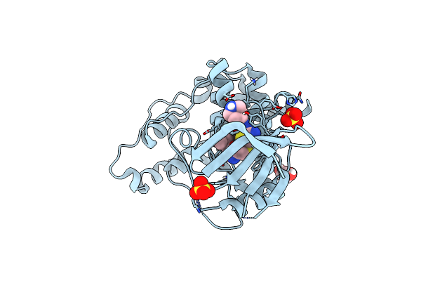 |
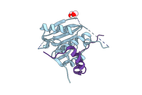
Organism: Homo sapiens
Method: X-RAY DIFFRACTION Resolution:2.60 Å Release Date: 2025-04-02 Classification: GENE REGULATION Ligands: A1H6P, SO4, GOL, PEG |
 |
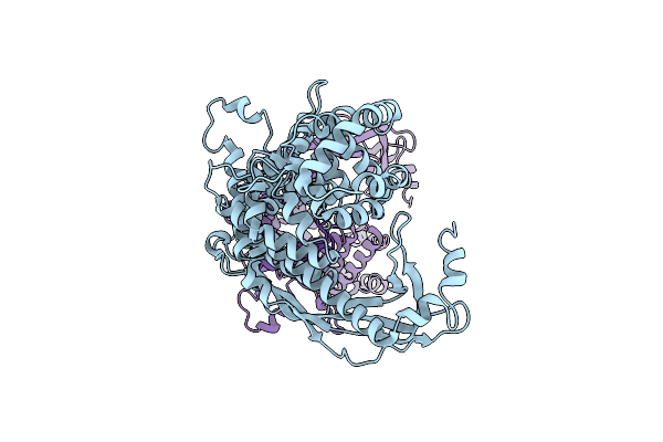
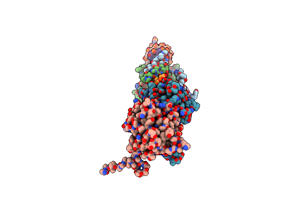
Organism: Leishmania tarentolae
Method: ELECTRON MICROSCOPY Release Date: 2025-03-12 Classification: STRUCTURAL PROTEIN Ligands: GDP, MG, GTP, ZN, CA, ATP |
 |
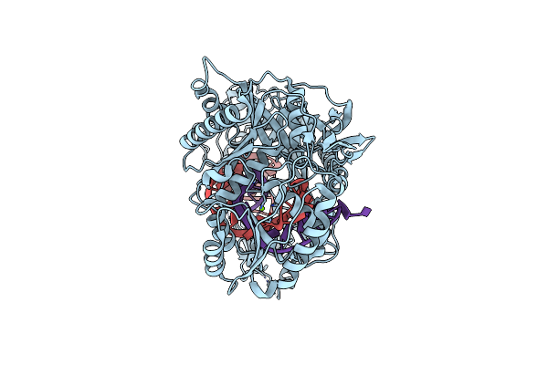
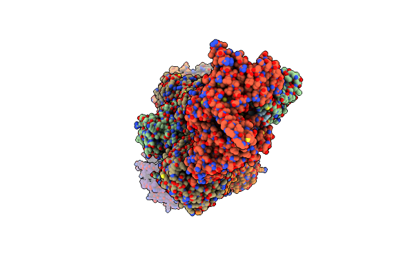
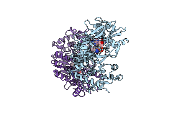
P116 Dimer In The Full State (Pdb Structure Of The Full-Length Ectodomain Truncated To Amino Acids 246-818)
Organism: Mycoplasmoides pneumoniae m129
Method: ELECTRON MICROSCOPY Release Date: 2024-05-29 Classification: LIPID BINDING PROTEIN |
 |
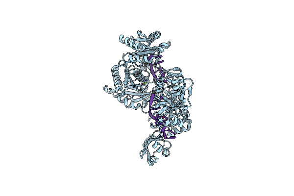
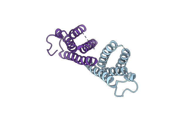
Organism: Ephydatia fluviatilis, Homo sapiens
Method: ELECTRON MICROSCOPY Release Date: 2024-02-14 Classification: RNA BINDING PROTEIN/RNA Ligands: MG |
 |
Organism: Homo sapiens
Method: ELECTRON MICROSCOPY Release Date: 2024-01-24 Classification: RNA BINDING PROTEIN/RNA Ligands: MG |
 |
Organism: Mus musculus, Homo sapiens
Method: ELECTRON MICROSCOPY Release Date: 2024-01-24 Classification: RNA BINDING PROTEIN/RNA Ligands: MG |
 |
Organism: Ephydatia fluviatilis, Homo sapiens
Method: ELECTRON MICROSCOPY Release Date: 2024-01-24 Classification: RNA BINDING PROTEIN/RNA Ligands: MG |
 |
Organism: Mus musculus, Homo sapiens
Method: ELECTRON MICROSCOPY Release Date: 2024-01-24 Classification: RNA BINDING PROTEIN/RNA Ligands: MG |
 |
Organism: Leishmania tarentolae
Method: ELECTRON MICROSCOPY Release Date: 2022-12-21 Classification: PROTEIN TRANSPORT Ligands: ZN |
 |
Organism: Leishmania tarentolae
Method: ELECTRON MICROSCOPY Release Date: 2022-12-21 Classification: PROTEIN TRANSPORT Ligands: ZN |
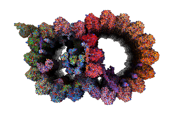 |
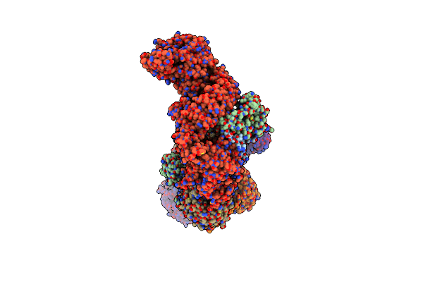
Structure Of The 48-Nm Repeat Doublet Microtubule From Bovine Tracheal Cilia
Organism: Bos taurus
Method: ELECTRON MICROSCOPY Release Date: 2021-10-27 Classification: STRUCTURAL PROTEIN Ligands: GTP, MG, GDP |
 |
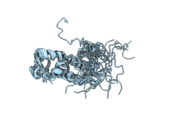
Organism: Legionella pneumophila
Method: SOLUTION NMR Release Date: 2020-07-22 Classification: UNKNOWN FUNCTION |
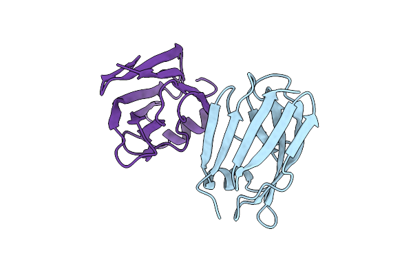 |
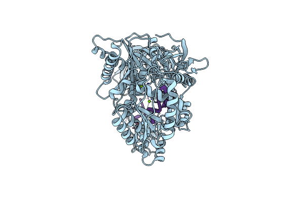
Crystal Structure Of The Legionella Pneumophila Type Ii Secretion System Substrate Nttc
Organism: Legionella pneumophila 130b
Method: X-RAY DIFFRACTION Resolution:3.10 Å Release Date: 2020-05-20 Classification: UNKNOWN FUNCTION Ligands: HOH |
 |
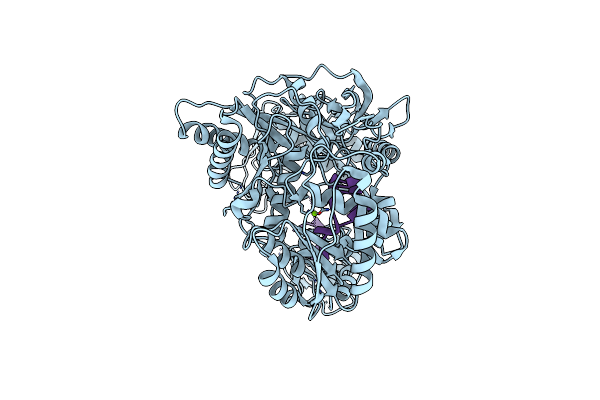
Crystal Structure Of The Legionella Pneumophila Type Ii Secretion System Substrate Ntte
Organism: Legionella pneumophila 130b
Method: X-RAY DIFFRACTION Resolution:2.20 Å Release Date: 2020-05-20 Classification: UNKNOWN FUNCTION Ligands: EDO |
 |
 |
Organism: Leishmania major
Method: X-RAY DIFFRACTION Resolution:2.70 Å Release Date: 2018-03-14 Classification: TRANSLATION Ligands: PEG |
 |
Organism: Bacillus subtilis, Synthetic construct
Method: X-RAY DIFFRACTION Resolution:3.21 Å Release Date: 2016-12-07 Classification: HYDROLASE |
 |
Organism: Herbaspirillum seropedicae
Method: X-RAY DIFFRACTION Resolution:1.70 Å Release Date: 2016-08-03 Classification: DNA BINDING PROTEIN Ligands: ADP, ATP, CA |
 |
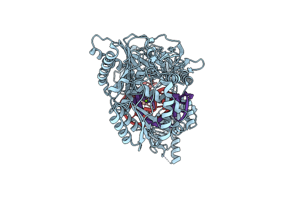
Crystal Structure Of The Anthranilate Coa Ligase Auaeii In Complex With Anthranoyl-Amp
Organism: Stigmatella aurantiaca
Method: X-RAY DIFFRACTION Resolution:2.60 Å Release Date: 2015-11-11 Classification: ligase/ligase inhibitor Ligands: 3UK |
 |
Organism: Mus musculus
Method: X-RAY DIFFRACTION Resolution:2.06 Å Release Date: 2014-09-03 Classification: PROTEIN BINDING |