
Search Count: 40
 |
Organism: Rocahepevirus ratti, Homo sapiens
Method: ELECTRON MICROSCOPY Release Date: 2025-07-23 Classification: VIRAL PROTEIN/IMMUNE SYSTEM |
 |
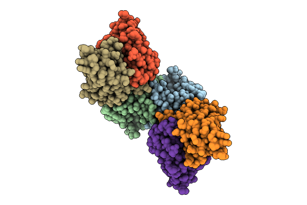
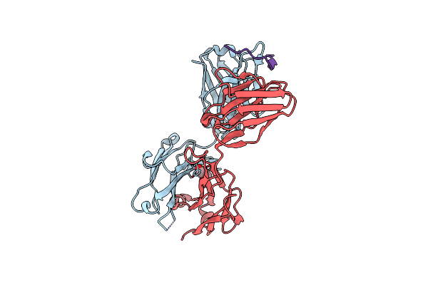
Hepatitis E Virus Capsid Protein E2S Domain (Genotype I) In Complex With Fab C6
Organism: Hepatitis e virus genotype 1 (isolate human/burma), Homo sapiens
Method: ELECTRON MICROSCOPY Release Date: 2025-07-23 Classification: VIRAL PROTEIN/IMMUNE SYSTEM |
 |
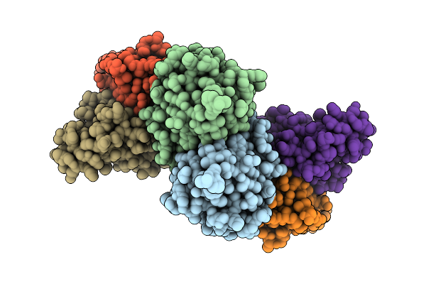
Hepatitis E Virus Capsid Protein E2S Domain (Genotype Iv) In Complex With Fab C6
Organism: Hepatitis e virus, Homo sapiens
Method: ELECTRON MICROSCOPY Release Date: 2025-07-23 Classification: VIRAL PROTEIN/IMMUNE SYSTEM |
 |
Organism: Rocahepevirus ratti, Homo sapiens
Method: ELECTRON MICROSCOPY Release Date: 2025-07-23 Classification: VIRAL PROTEIN/IMMUNE SYSTEM |
 |
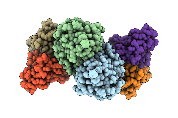
Hepatitis E Virus Capsid Protein E2S Domain (Genotype I) In Complex With Fab C158
Organism: Hepatitis e virus genotype 1 (isolate human/burma), Homo sapiens
Method: ELECTRON MICROSCOPY Release Date: 2025-07-23 Classification: VIRAL PROTEIN/IMMUNE SYSTEM |
 |
Organism: Rocahepevirus ratti, Homo sapiens
Method: ELECTRON MICROSCOPY Release Date: 2025-07-23 Classification: VIRAL PROTEIN/IMMUNE SYSTEM |
 |
Organism: Rocahepevirus ratti, Homo sapiens
Method: ELECTRON MICROSCOPY Release Date: 2025-07-23 Classification: VIRAL PROTEIN/IMMUNE SYSTEM |
 |
Organism: Rocahepevirus ratti, Homo sapiens
Method: ELECTRON MICROSCOPY Release Date: 2025-07-23 Classification: VIRAL PROTEIN/IMMUNE SYSTEM |
 |
Organism: Rocahepevirus ratti, Homo sapiens
Method: ELECTRON MICROSCOPY Release Date: 2025-07-23 Classification: VIRAL PROTEIN/IMMUNE SYSTEM |
 |
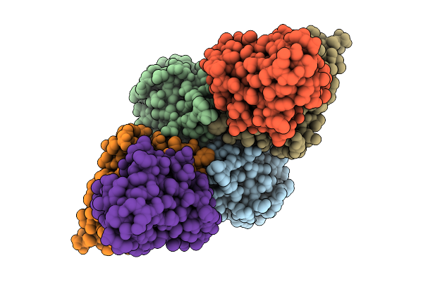
Hepatitis E Virus Capsid Protein E2S Domain (Genotype I) In Complex With Fab H4
Organism: Hepatitis e virus genotype 1 (isolate human/burma), Homo sapiens
Method: ELECTRON MICROSCOPY Release Date: 2025-07-23 Classification: VIRAL PROTEIN/IMMUNE SYSTEM |
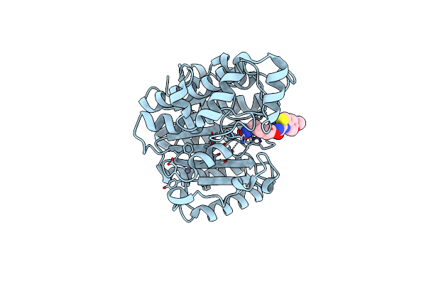 |
Crystal Structure Of Danio Rerio Histone Deacetylase 6 Catalytic Domain 2 (Cd2) In Complex With N-((6-(Hydroxyamino)-6-Oxohexyl)Oxy)-4-(4-Methoxyphenyl)Thiazole-2-Carboxamide
Organism: Danio rerio
Method: X-RAY DIFFRACTION Resolution:1.90 Å Release Date: 2024-12-11 Classification: HYDROLASE Ligands: A1IK2, ZN |
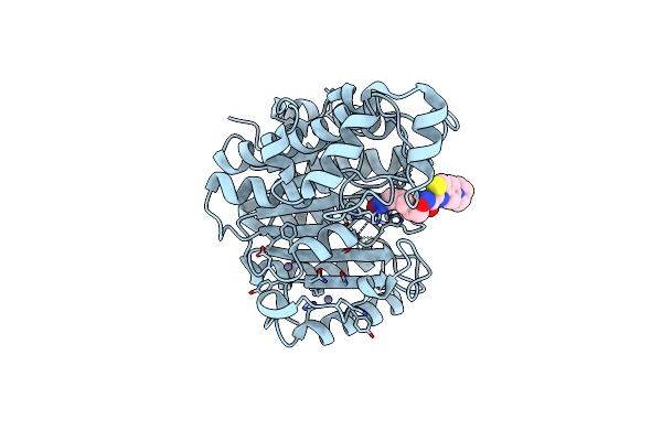 |
Crystal Structure Of Danio Rerio Histone Deacetylase 6 Catalytic Domain 2 (Cd2) In Complex With N-((6-(Hydroxyamino)-6-Oxohexyl)Oxy)-4-(4-(Pyrrolidin-1-Yl)Phenyl)Thiazole-2-Carboxamide
Organism: Danio rerio
Method: X-RAY DIFFRACTION Resolution:2.14 Å Release Date: 2024-12-04 Classification: HYDROLASE Ligands: A1IKY, ZN |
 |
Organism: Homo sapiens, Zika virus
Method: ELECTRON MICROSCOPY Release Date: 2022-11-23 Classification: VIRUS |
 |
Organism: Homo sapiens, Human betaherpesvirus 5
Method: X-RAY DIFFRACTION Resolution:1.80 Å Release Date: 2022-10-19 Classification: IMMUNE SYSTEM/VIRAL PROTEIN Ligands: GOL |
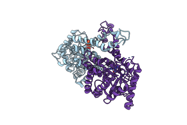 |
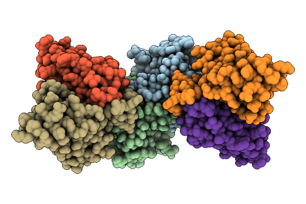
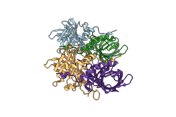
Structure Of Candida Albicans Fructose-1,6-Bisphosphate Aldolase Complexed With G3P
Organism: Candida albicans sc5314
Method: X-RAY DIFFRACTION Resolution:2.98 Å Release Date: 2022-02-23 Classification: LYASE Ligands: ZN, G3H |
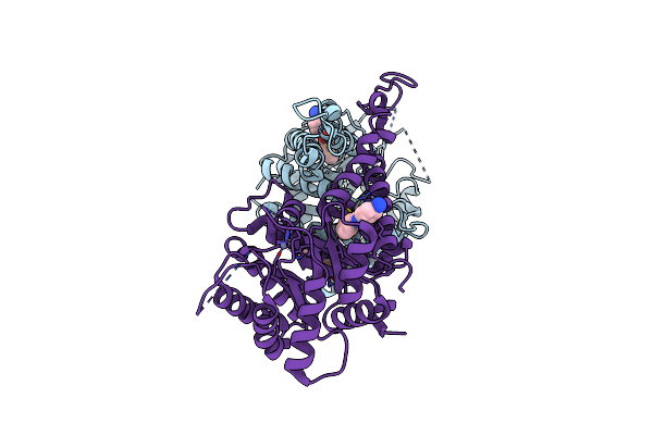 |
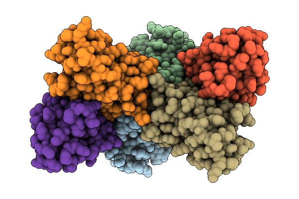
Structure Of Candida Albicans Fructose-1,6-Bisphosphate Aldolase Mutation C157S With Cn39
Organism: Candida albicans sc5314
Method: X-RAY DIFFRACTION Resolution:2.34 Å Release Date: 2022-02-23 Classification: LYASE Ligands: ZN, EDO, 6Y3 |
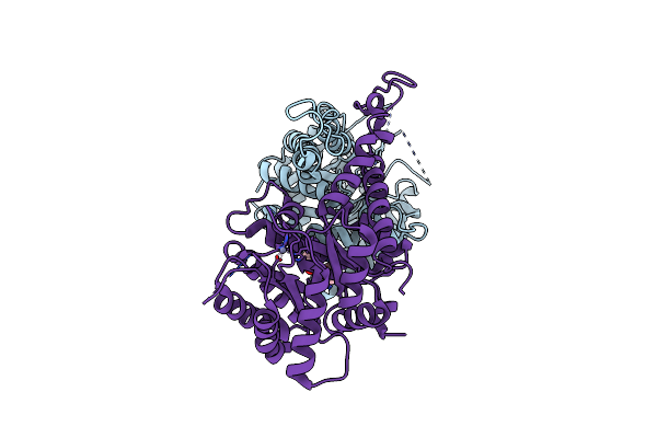 |
Organism: Candida albicans sc5314
Method: X-RAY DIFFRACTION Resolution:2.64 Å Release Date: 2020-12-30 Classification: LYASE Ligands: ZN, EDO |
 |
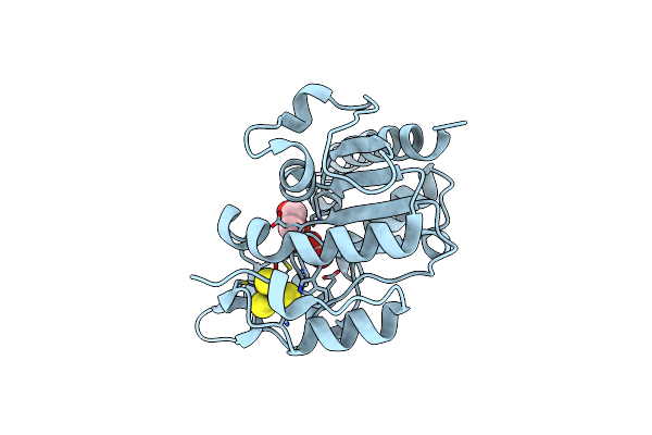
A Nonspecific Heme-Binding Cyclase Catalyzes [4 + 2] Cycloaddition During Neoabyssomicin Biosynthesis
Organism: Streptomyces koyangensis
Method: X-RAY DIFFRACTION Resolution:2.51 Å Release Date: 2020-11-25 Classification: BIOSYNTHETIC PROTEIN |
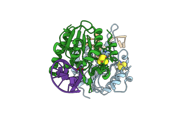 |
Organism: Mycolicibacterium smegmatis mc2 155, Synthetic construct
Method: X-RAY DIFFRACTION Resolution:2.06 Å Release Date: 2020-11-04 Classification: DNA BINDING PROTEIN Ligands: SF4 |
 |
Organism: Mycolicibacterium smegmatis mc2 155
Method: X-RAY DIFFRACTION Resolution:1.80 Å Release Date: 2020-10-28 Classification: DNA BINDING PROTEIN Ligands: SF4, GOL |