
Search Count: 39
 |
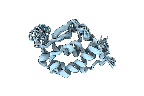
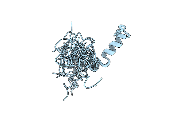
Nmr Structure Of The Staphylococcus Aureus Bacteriophage Phi812 Hub Protein - Lytic Cleaver (Chap) Domain
Organism: Staphylococcus phage 812
Method: SOLUTION NMR Release Date: 2025-04-09 Classification: HYDROLASE |
 |
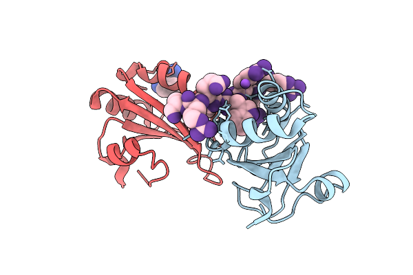
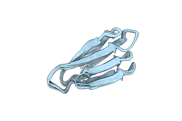
X-Ray Structure Of Dishevelled 3 Pdz Domain In A Complex With A Class Iii Peptide Ligand
Organism: Homo sapiens
Method: X-RAY DIFFRACTION Release Date: 2025-03-12 Classification: SIGNALING PROTEIN |
 |
Organism: Homo sapiens
Method: X-RAY DIFFRACTION Resolution:2.49 Å Release Date: 2022-12-28 Classification: CYTOKINE |
 |
Organism: Homo sapiens
Method: X-RAY DIFFRACTION Resolution:3.34 Å Release Date: 2022-12-28 Classification: CYTOKINE Ligands: SO4 |
 |
Organism: Homo sapiens
Method: X-RAY DIFFRACTION Resolution:1.70 Å Release Date: 2022-12-28 Classification: CYTOKINE Ligands: SO4 |
 |
Organism: Mus musculus
Method: X-RAY DIFFRACTION Resolution:1.80 Å Release Date: 2022-12-28 Classification: CYTOKINE Ligands: ZN, ACT |
 |
Organism: Homo sapiens
Method: X-RAY DIFFRACTION Resolution:3.09 Å Release Date: 2022-12-28 Classification: CYTOKINE Ligands: SO4 |
 |
 |
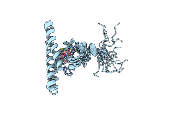
Crystal Structure Of The N-Terminal Ef-Hand Domain Of Arabidopsis Thaliana Ateh1/Pan1
Organism: Arabidopsis thaliana
Method: X-RAY DIFFRACTION Resolution:1.55 Å Release Date: 2021-04-14 Classification: ENDOCYTOSIS Ligands: CA, NA |
 |
Organism: Arabidopsis thaliana
Method: SOLUTION NMR Release Date: 2021-03-31 Classification: LIPID BINDING PROTEIN Ligands: CA |
 |
Organism: Arabidopsis thaliana
Method: SOLUTION NMR Release Date: 2021-03-31 Classification: PEPTIDE BINDING PROTEIN Ligands: CA |
 |
Organism: Homo sapiens, Human immunodeficiency virus 1
Method: X-RAY DIFFRACTION Resolution:1.20 Å Release Date: 2019-03-27 Classification: SPLICING Ligands: TRP, NA |
 |
Organism: Synthetic construct
Method: SOLUTION NMR Release Date: 2018-11-07 Classification: DE NOVO PROTEIN |
 |
Organism: Lysobacter enzymogenes
Method: SOLUTION NMR Release Date: 2018-02-07 Classification: HYDROLASE |
 |
Organism: Mycobacterium smegmatis (strain atcc 700084 / mc(2)155)
Method: SOLUTION NMR Release Date: 2018-02-07 Classification: UNKNOWN FUNCTION |
 |
Organism: Caldanaerobacter subterraneus subsp. tengcongensis (strain dsm 15242 / jcm 11007 / nbrc 100824 / mb4)
Method: SOLUTION NMR Release Date: 2018-02-07 Classification: TRANSFERASE |
 |
Organism: Saccharomyces cerevisiae (strain atcc 204508 / s288c)
Method: SOLUTION NMR Release Date: 2018-02-07 Classification: TRANSCRIPTION REGULATOR |
 |
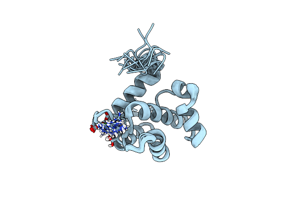
Solution Structure Of Oxidized And Amidated Human Iapp (1-37), The Diabetes Ii Peptide.
|
 |
Organism: Homo sapiens
Method: SOLUTION NMR Release Date: 2017-03-15 Classification: CHAPERONE Ligands: FAR |
 |
Targeting The Pex14-Pex5 Interaction By Small Molecules Provides Novel Therapeutic Routes To Treat Trypanosomiases.
Organism: Trypanosoma brucei brucei
Method: X-RAY DIFFRACTION Resolution:1.55 Å Release Date: 2017-03-15 Classification: SIGNALING PROTEIN Ligands: KZZ, SO4, BME, CL |