
Search Count: 38
 |
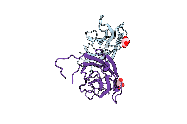
Crystal Structure Of A Calcium Bound C2 Domain Containing Protein From Trichomonas Vaginalis
Organism: Trichomonas vaginalis g3
Method: X-RAY DIFFRACTION Release Date: 2025-12-17 Classification: LIPID BINDING PROTEIN Ligands: 1PE, CL, PEG, PGE |
 |
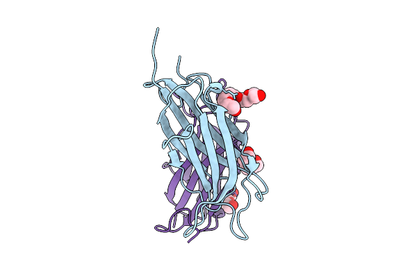
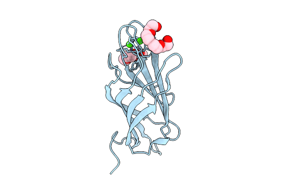
Crystal Structure Of A Calcium Bound C2 Domain Containing Protein From Trichomonas Vaginalis (P21 Form)
Organism: Trichomonas vaginalis g3
Method: X-RAY DIFFRACTION Release Date: 2025-12-17 Classification: LIPID BINDING PROTEIN Ligands: PG4, CA, MES, 1PE |
 |
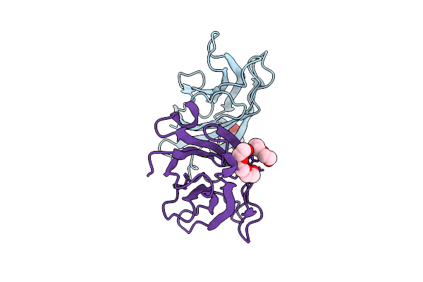
Crystal Structure Of A Calcium Bound C2 Domain Containing Protein From Trichomonas Vaginalis (Orthorhombic P Form)
Organism: Trichomonas vaginalis g3
Method: X-RAY DIFFRACTION Release Date: 2025-12-17 Classification: LIPID BINDING PROTEIN Ligands: CA, 1PE |
 |
Crystal Structure Of A C2 Domain Containing Protein From Trichomonas Vaginalis
Organism: Trichomonas vaginalis g3
Method: X-RAY DIFFRACTION Release Date: 2025-12-17 Classification: LIPID BINDING PROTEIN Ligands: GOL, PGE |
 |
Crystal Structure Of A Malonate Bound C2 Domain Containing Protein From Trichomonas Vaginalis (P21 Form)
Organism: Trichomonas vaginalis g3
Method: X-RAY DIFFRACTION Release Date: 2025-12-17 Classification: LIPID BINDING PROTEIN Ligands: MLI, CL, NA |
 |
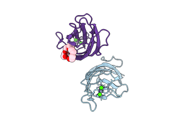
Crystal Structure Of A Calcium Bound C2 Domain Containing Protein From Trichomonas Vaginalis (P61 Form)
Organism: Trichomonas vaginalis g3
Method: X-RAY DIFFRACTION Release Date: 2025-12-17 Classification: LIPID BINDING PROTEIN Ligands: CA, CL, PG4 |
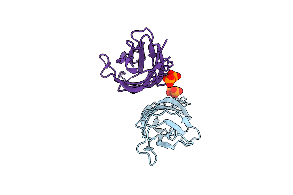 |
Crystal Structure Of A C2 Domain Containing Protein From Trichomonas Vaginalis In Complex With Pyrophosphate
Organism: Trichomonas vaginalis g3
Method: X-RAY DIFFRACTION Release Date: 2025-12-17 Classification: LIPID BINDING PROTEIN Ligands: PPV |
 |
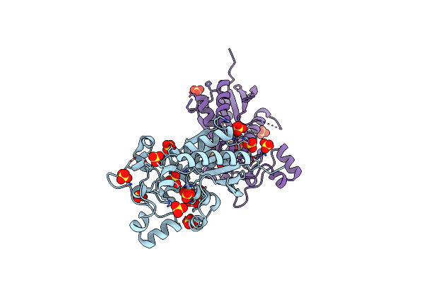
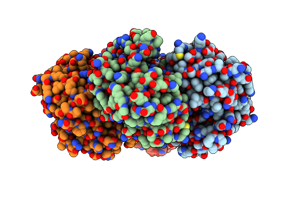
Crystal Structure Of Phosphoglycerate Mutase From Trichomonas Vaginalis In Complex With 3-Phosphoglyceric Acid
Organism: Trichomonas vaginalis g3
Method: X-RAY DIFFRACTION Release Date: 2025-12-03 Classification: ISOMERASE Ligands: CL, 3PG, PG4, SO4 |
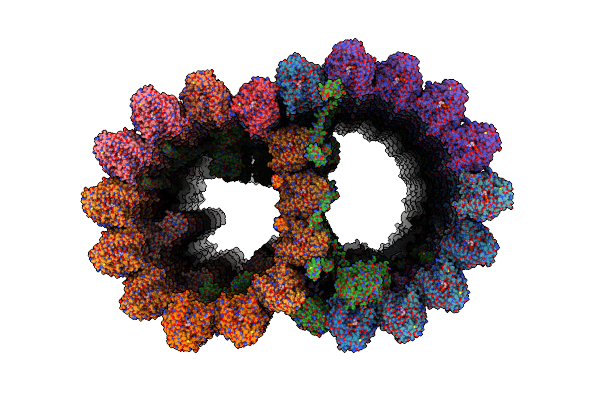 |
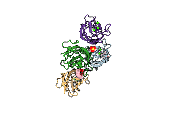
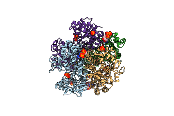
Organism: Trichomonas vaginalis g3
Method: ELECTRON MICROSCOPY Release Date: 2025-05-14 Classification: STRUCTURAL PROTEIN Ligands: GDP, GTP, MG |
 |
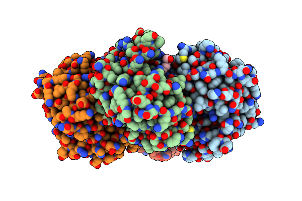
Crystal Structure Of Phosphoglycerate Mutase From Trichomonas Vaginalis (Sulfate Bound)
Organism: Trichomonas vaginalis g3
Method: X-RAY DIFFRACTION Resolution:2.05 Å Release Date: 2025-04-23 Classification: ISOMERASE Ligands: SO4 |
 |
Organism: Trichomonas vaginalis g3
Method: X-RAY DIFFRACTION Resolution:1.95 Å Release Date: 2025-04-02 Classification: ISOMERASE Ligands: SO4, GOL, P33, PG4 |
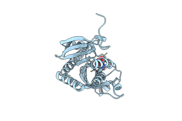 |
Crystal Structure Of Purine Nucleoside Phosphorylase From Trichomonas Vaginalis (Inosine Bound)
Organism: Trichomonas vaginalis g3
Method: X-RAY DIFFRACTION Resolution:1.20 Å Release Date: 2025-01-22 Classification: TRANSFERASE Ligands: NOS, CL |
 |
Crystal Structure Of Purine Nucleoside Phosphorylase From Trichomonas Vaginalis (C2 Form)
Organism: Trichomonas vaginalis g3
Method: X-RAY DIFFRACTION Resolution:1.80 Å Release Date: 2024-12-25 Classification: TRANSFERASE Ligands: GOL |
 |
Crystal Structure Of Purine Nucleoside Phosphorylase From Trichomonas Vaginalis (P21 Form)
Organism: Trichomonas vaginalis g3
Method: X-RAY DIFFRACTION Resolution:1.59 Å Release Date: 2024-12-25 Classification: TRANSFERASE Ligands: FLC |
 |
Crystal Structure Of Purine Nucleoside Phosphorylase From Trichomonas Vaginalis (Hepes Bound)
Organism: Trichomonas vaginalis g3
Method: X-RAY DIFFRACTION Resolution:1.49 Å Release Date: 2024-12-25 Classification: TRANSFERASE Ligands: GOL, EPE |
 |
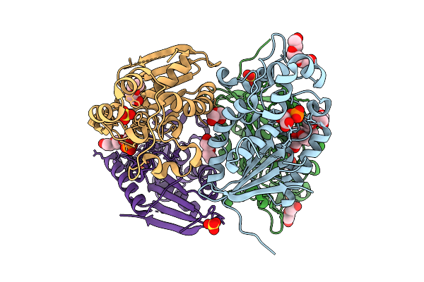
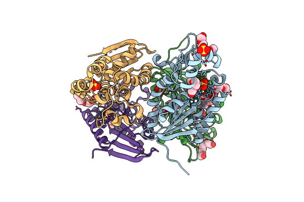
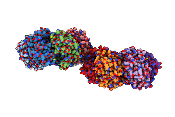
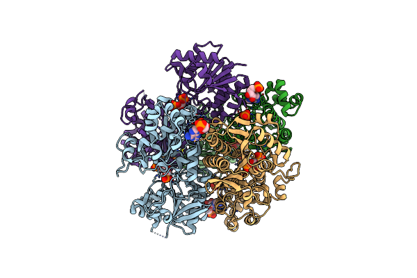
Crystal Structure Pyrophosphate-Fructose 6-Phosphate 1-Phosphotransferase 1 (Pfk1) From Trichomonas Vaginalis (Atp/Pyrophosphate Complex)
Organism: Trichomonas vaginalis g3
Method: X-RAY DIFFRACTION Resolution:2.16 Å Release Date: 2024-12-18 Classification: TRANSFERASE Ligands: ATP, PPV, AMP, PO4, CL |
 |
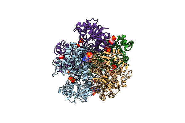
Crystal Structure Of Pyrophosphate-Fructose 6-Phosphate 1-Phosphotransferase 1 (Pfk1) From Trichomonas Vaginalis (Amp/Fructose-1,6-Biphosphate Complex)
Organism: Trichomonas vaginalis g3
Method: X-RAY DIFFRACTION Resolution:2.83 Å Release Date: 2024-12-18 Classification: TRANSFERASE Ligands: FBP, PO4, AMP |
 |
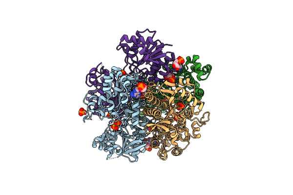
Crystal Structure Of Pyrophosphate-Fructose 6-Phosphate 1-Phosphotransferase 1 (Pfk1) From Trichomonas Vaginalis (Amp/Alpha-D-Glucose-6-Phosphate Complex)
Organism: Trichomonas vaginalis g3
Method: X-RAY DIFFRACTION Resolution:2.80 Å Release Date: 2024-12-18 Classification: TRANSFERASE Ligands: AMP, PPV, GOL, G6P, PO4, CL, MG |
 |
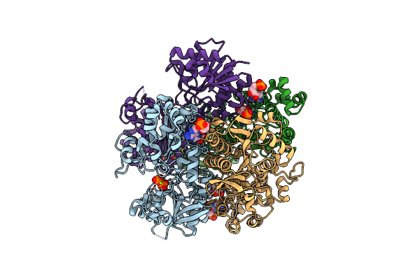
Crystal Structure Pyrophosphate-Fructose 6-Phosphate 1-Phosphotransferase 1 (Pfk1) From Trichomonas Vaginalis (Amp/Beta-D-Glucose-6-Phosphate Complex)
Organism: Trichomonas vaginalis g3
Method: X-RAY DIFFRACTION Resolution:2.62 Å Release Date: 2024-12-18 Classification: TRANSFERASE Ligands: PO4, AMP, PPV, MG, BG6, CL |
 |
Crystal Structure Of Pyrophosphate-Fructose 6-Phosphate 1-Phosphotransferase 1 (Pfk1) From Trichomonas Vaginalis (Adp/5-O-Phosphono-Alpha-D-Ribofuranose Complex)
Organism: Trichomonas vaginalis g3
Method: X-RAY DIFFRACTION Resolution:2.65 Å Release Date: 2024-12-18 Classification: TRANSFERASE Ligands: HSX, AMP, MG, PPV |