
Search Count: 25
 |
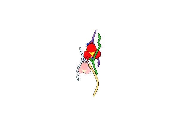
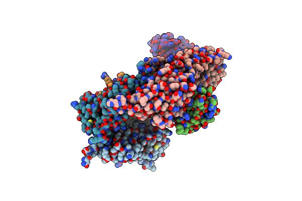
Structure Of Ecarin From The Venom Of Kenyan Saw-Scaled Viper In Complex With The Fab Of Neutralizing Antibody H11
Organism: Echis carinatus, Homo sapiens
Method: ELECTRON MICROSCOPY Release Date: 2024-09-11 Classification: HYDROLASE Ligands: ZN, CA |
 |
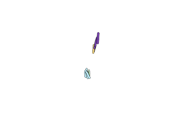
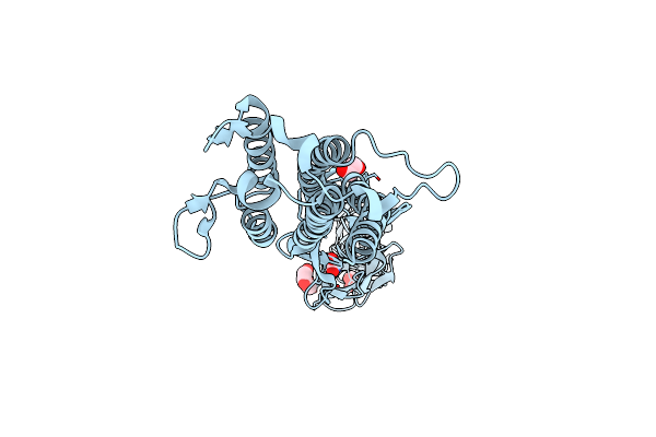
Crystal Structure Of Long Neurotoxin From The Venom Of The King Cobra (3Ftx-L15) In Complex With Fab Of Broadly Neutralizing Antibody 95Mat5
Organism: Homo sapiens, Ophiophagus hannah
Method: X-RAY DIFFRACTION Resolution:2.90 Å Release Date: 2024-03-20 Classification: TOXIN/IMMUNE SYSTEM |
 |
Crystal Structure Of Sars-Cov-2 Receptor Binding Domain In Complex With Antibodies Ecr3022.20 And Cc12.3
Organism: Severe acute respiratory syndrome coronavirus 2, Homo sapiens
Method: X-RAY DIFFRACTION Resolution:2.85 Å Release Date: 2023-08-23 Classification: IMMUNE SYSTEM/VIRAL PROTEIN Ligands: NAG |
 |
Crystal Structure Of Sars-Cov-2 Spike Protein Receptor-Binding Domain In Complex With Antibody Cc12.1 Fab And Nanobody Nb-C4-225
Organism: Severe acute respiratory syndrome coronavirus 2, Synthetic construct, Homo sapiens
Method: X-RAY DIFFRACTION Resolution:2.72 Å Release Date: 2023-07-05 Classification: VIRAL PROTEIN/IMMUNE SYSTEM Ligands: NAG |
 |
Crystal Structure Of Sars-Cov-2 Spike Protein Receptor-Binding Domain In Complex With Antibody Cc12.1 Fab And Nanobody Nb-C4-240
Organism: Severe acute respiratory syndrome coronavirus 2, Synthetic construct, Homo sapiens
Method: X-RAY DIFFRACTION Resolution:2.83 Å Release Date: 2023-07-05 Classification: VIRAL PROTEIN/IMMUNE SYSTEM Ligands: NAG |
 |
Crystal Structure Of Sars-Cov-2 Spike Protein Receptor-Binding Domain In Complex With Antibody Cc12.1 Fab And Nanobody Nb-C4-255
Organism: Severe acute respiratory syndrome coronavirus 2, Synthetic construct, Homo sapiens
Method: X-RAY DIFFRACTION Resolution:2.21 Å Release Date: 2023-07-05 Classification: VIRAL PROTEIN/IMMUNE SYSTEM Ligands: NAG |
 |
Lm18/Nb136 Bispecific Tetra-Nanobody Immunoglobulin In Complex With Sars-Cov-2-6P-Mut7 S Protein (Focused Refinement)
Organism: Severe acute respiratory syndrome coronavirus 2, Synthetic construct
Method: ELECTRON MICROSCOPY Release Date: 2023-06-14 Classification: VIRAL PROTEIN Ligands: NAG |
 |
Organism: Severe acute respiratory syndrome coronavirus 2
Method: ELECTRON MICROSCOPY Release Date: 2022-08-24 Classification: VIRAL PROTEIN Ligands: NAG |
 |
Organism: Severe acute respiratory syndrome coronavirus 2
Method: ELECTRON MICROSCOPY Release Date: 2022-08-24 Classification: VIRAL PROTEIN Ligands: NAG |
 |
Cc6.33 Igg In Complex With Sars-Cov-2-6P-Mut7 S Protein (Non-Uniform Refinement)
Organism: Homo sapiens, Severe acute respiratory syndrome coronavirus 2
Method: ELECTRON MICROSCOPY Release Date: 2022-08-24 Classification: VIRAL PROTEIN/Immune System Ligands: NAG |
 |
Cc6.33 Igg In Complex With Sars-Cov-2-6P-Mut7 S Protein (Rbd/Fv Local Refinement)
Organism: Homo sapiens, Severe acute respiratory syndrome coronavirus 2
Method: ELECTRON MICROSCOPY Release Date: 2022-08-24 Classification: VIRAL PROTEIN/Immune System Ligands: NAG |
 |
Cc6.30 Fragment Antigen Binding In Complex With Sars-Cov-2-6P-Mut7 S Protein (Non-Uniform Refinement)
Organism: Severe acute respiratory syndrome coronavirus 2, Homo sapiens
Method: ELECTRON MICROSCOPY Release Date: 2022-08-24 Classification: VIRAL PROTEIN/Immune System Ligands: NAG |
 |
Cc6.30 Fragment Antigen Binding In Complex With Sars-Cov-2-6P-Mut7 S Protein (Rbd/Fv Local Refinement)
Organism: Homo sapiens, Severe acute respiratory syndrome coronavirus 2
Method: ELECTRON MICROSCOPY Release Date: 2022-08-24 Classification: VIRAL PROTEIN/Immune System Ligands: NAG |
 |
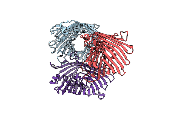
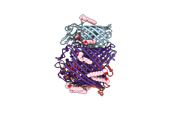
Structure Of 283-Lgny-286, The Steric Zipper That Supports The Self-Association Of P. Stuartii Omp-Pst2 Into Dimers Of Trimers
Organism: Providencia stuartii
Method: X-RAY DIFFRACTION Resolution:1.00 Å Release Date: 2018-02-21 Classification: CELL ADHESION Ligands: SO4 |
 |
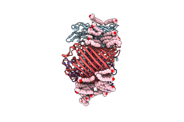
Structure Of 206-Gvvtse-211, The Steric Zipper That Supports The Self-Association Of P. Stuartii Omp-Pst1 Into Dimers Of Trimers
Organism: Providencia stuartii
Method: X-RAY DIFFRACTION Resolution:1.91 Å Release Date: 2018-02-21 Classification: CELL ADHESION |
 |
Organism: Providencia stuartii
Method: X-RAY DIFFRACTION Resolution:3.12 Å Release Date: 2018-02-21 Classification: TRANSPORT PROTEIN Ligands: CA |
 |
Organism: Providencia stuartii
Method: X-RAY DIFFRACTION Resolution:2.70 Å Release Date: 2018-02-21 Classification: CELL ADHESION Ligands: LDA, CA, CL |
 |
Organism: Providencia stuartii
Method: X-RAY DIFFRACTION Resolution:3.00 Å Release Date: 2018-02-21 Classification: CELL ADHESION Ligands: LDA, CA |
 |
Organism: Porphyromonas gingivalis, Lama glama
Method: X-RAY DIFFRACTION Resolution:2.40 Å Release Date: 2018-02-07 Classification: PROTEIN TRANSPORT |
 |
Organism: Flavobacterium johnsoniae
Method: X-RAY DIFFRACTION Resolution:2.00 Å Release Date: 2018-02-07 Classification: PROTEIN TRANSPORT Ligands: EDO |