
Search Count: 28
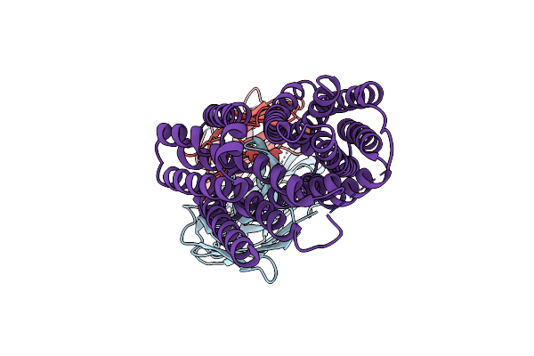 |
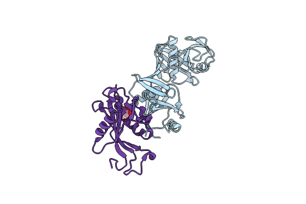
Organism: Staphylococcus aureus, Homo sapiens
Method: ELECTRON MICROSCOPY Release Date: 2024-12-11 Classification: TRANSPORT PROTEIN/IMMUNE SYSTEM |
 |
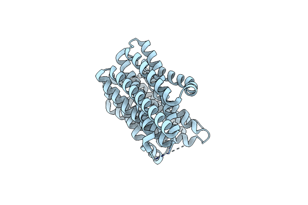
Organism: Staphylococcus aureus
Method: ELECTRON MICROSCOPY Release Date: 2024-12-11 Classification: TRANSPORT PROTEIN |
 |
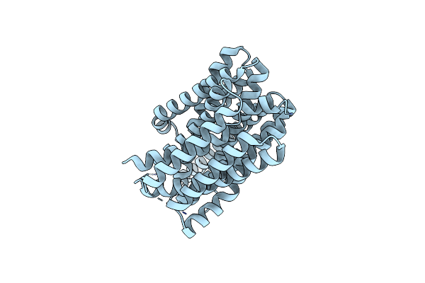
Organism: Staphylococcus aureus
Method: ELECTRON MICROSCOPY Release Date: 2024-12-11 Classification: TRANSPORT PROTEIN |
 |
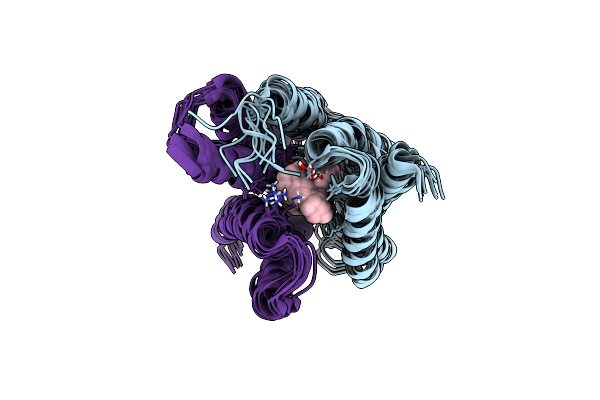
Organism: Staphylococcus aureus
Method: ELECTRON MICROSCOPY Release Date: 2024-12-11 Classification: TRANSPORT PROTEIN |
 |
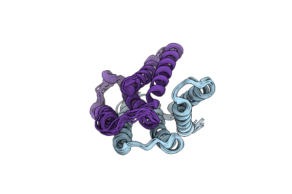
Organism: Staphylococcus aureus, Homo sapiens
Method: ELECTRON MICROSCOPY Release Date: 2024-05-29 Classification: TRANSPORT PROTEIN |
 |
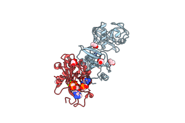
Organism: Staphylococcus aureus, Homo sapiens
Method: ELECTRON MICROSCOPY Release Date: 2024-05-29 Classification: TRANSPORT PROTEIN/IMMUNE SYSTEM |
 |
Organism: Staphylococcus aureus, Homo sapiens
Method: ELECTRON MICROSCOPY Release Date: 2024-05-29 Classification: TRANSPORT PROTEIN |
 |
Organism: Staphylococcus aureus, Homo sapiens
Method: ELECTRON MICROSCOPY Release Date: 2024-05-29 Classification: TRANSPORT PROTEIN/IMMUNE SYSTEM |
 |
Organism: Escherichia coli
Method: SOLUTION NMR, SOLID-STATE NMR Release Date: 2024-05-29 Classification: MEMBRANE PROTEIN Ligands: P4P |
 |
Organism: Escherichia coli
Method: SOLUTION NMR, SOLID-STATE NMR Release Date: 2024-05-29 Classification: MEMBRANE PROTEIN |
 |
Organism: Staphylococcus aureus, Homo sapiens
Method: ELECTRON MICROSCOPY Release Date: 2022-04-20 Classification: TRANSPORT PROTEIN |
 |
Organism: Staphylococcus aureus, Homo sapiens
Method: ELECTRON MICROSCOPY Release Date: 2022-04-20 Classification: TRANSPORT PROTEIN |
 |
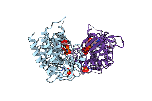
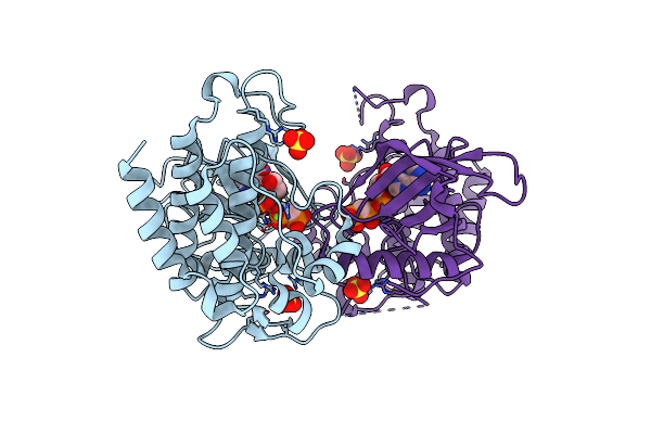
Crystal Structure Of An Asymmetric Dimer Of Fgf Receptor 3 Kinases Trapped In A-Loop Tyrosine Transphosphorylation Reaction
Organism: Homo sapiens
Method: X-RAY DIFFRACTION Resolution:2.20 Å Release Date: 2020-01-22 Classification: TRANSFERASE Ligands: ACP, SO4 |
 |
Organism: Escherichia coli o45:k1 (strain s88 / expec)
Method: X-RAY DIFFRACTION Resolution:3.06 Å Release Date: 2018-05-23 Classification: isomerase/hydrolase Ligands: IOD, CL |
 |
Organism: Escherichia coli (strain k12)
Method: X-RAY DIFFRACTION Resolution:2.15 Å Release Date: 2018-05-23 Classification: isomerase/hydrolase Ligands: CL, GOL |
 |
Organism: Escherichia coli
Method: X-RAY DIFFRACTION Resolution:1.81 Å Release Date: 2018-05-23 Classification: isomerase/hydrolase/rna Ligands: GOL, CL, MG |
 |
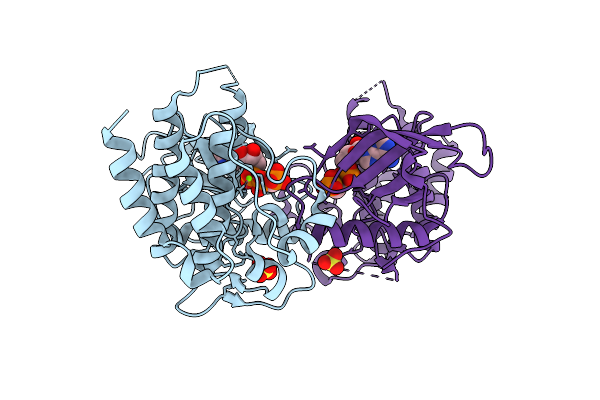
Crystal Structure Of Fgf Receptor 2 Tyrosine Kinase Domain Harboring The D650V Activating Mutation
Organism: Homo sapiens
Method: X-RAY DIFFRACTION Resolution:1.86 Å Release Date: 2017-02-22 Classification: TRANSFERASE Ligands: SO4, ANP |
 |
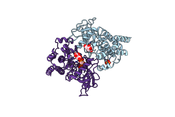
Crystal Structure Of The Tyrosine Kinase Domain Of Fgf Receptor 2 Harboring A E565A/D650V Double Gain-Of-Function Mutation
Organism: Homo sapiens
Method: X-RAY DIFFRACTION Resolution:2.35 Å Release Date: 2017-02-22 Classification: TRANSFERASE Ligands: SO4, ACP, MG |
 |
Crystal Structure Of The Tyrosine Kinase Domain Of Fgf Receptor 2 Harboring A N549H/E565A Double Gain-Of-Function Mutation
Organism: Homo sapiens
Method: X-RAY DIFFRACTION Resolution:2.91 Å Release Date: 2017-02-22 Classification: TRANSFERASE Ligands: SO4, ACP, MG |
 |
Crystal Structure Of The Tyrosine Kinase Domain Of Fgf Receptor 2 Harboring An E565A/K659M Double Gain-Of-Function Mutation
Organism: Homo sapiens
Method: X-RAY DIFFRACTION Resolution:2.05 Å Release Date: 2017-02-22 Classification: TRANSFERASE Ligands: FLC, SO4, ACP |