
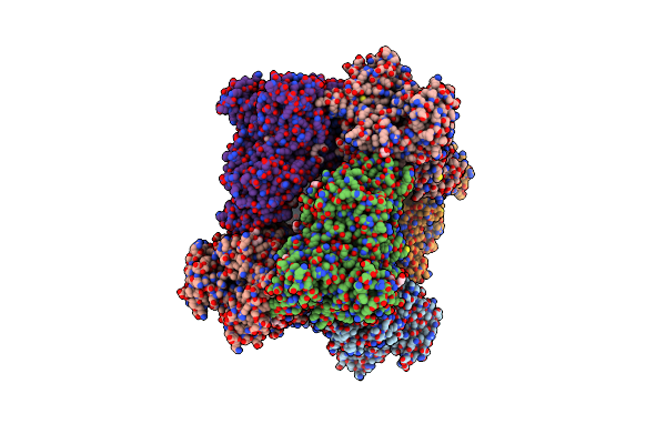
Search Count: 12
 |
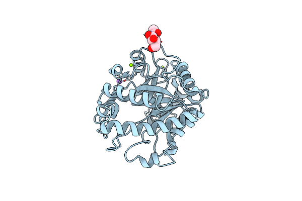
Crystal Structure Of Gdp-Bound Gnas In Complex With The Cyclic Peptide Inhibitor Gd20
Organism: Homo sapiens, Synthetic construct
Method: X-RAY DIFFRACTION Resolution:1.95 Å Release Date: 2022-03-02 Classification: SIGNALING PROTEIN Ligands: GDP, CL |
 |
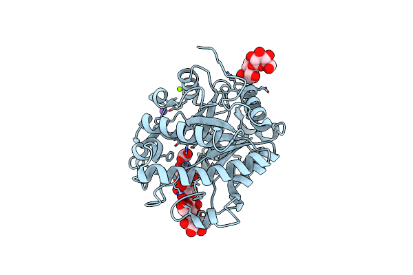
Crystal Structure Of Gppnhp-Bound Gnas In Complex With The Cyclic Peptide Inhibitor Gn13
Organism: Homo sapiens, Synthetic construct
Method: X-RAY DIFFRACTION Resolution:1.57 Å Release Date: 2021-03-31 Classification: SIGNALING PROTEIN Ligands: GNP, MG, GOL, CL |
 |
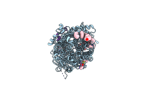
Cryo-Em Structure Of The G Protein-Gated Inward Rectifier K+ Channel Girk2 (Kir3.2) In Apo Form
Organism: Mus musculus
Method: ELECTRON MICROSCOPY Release Date: 2020-10-07 Classification: MEMBRANE PROTEIN |
 |
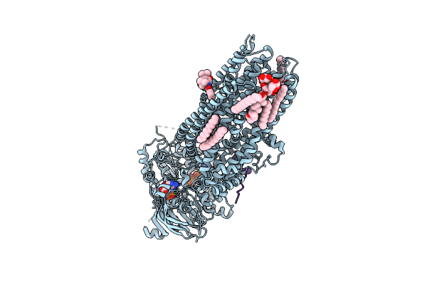
Cryo-Em Structure Of The G Protein-Gated Inward Rectifier K+ Channel Girk2 (Kir3.2) In Complex With Pip2
Organism: Mus musculus
Method: ELECTRON MICROSCOPY Release Date: 2020-10-07 Classification: MEMBRANE PROTEIN Ligands: PIO, K |
 |
Complex Of Human Cystic Fibrosis Transmembrane Conductance Regulator (Cftr) And Glpg1837
Organism: Homo sapiens
Method: ELECTRON MICROSCOPY Release Date: 2019-06-26 Classification: HYDROLASE Ligands: LJP, MG, ATP, POV, CLR |
 |
Complex Of Ivacaftor With Cystic Fibrosis Transmembrane Conductance Regulator (Cftr)
Organism: Homo sapiens
Method: ELECTRON MICROSCOPY Release Date: 2019-06-26 Classification: HYDROLASE Ligands: MG, ATP, POV, AJP, VX7 |
 |
Crystal Structure Of The P2X4 Receptor From Zebrafish In The Presence Of Ctp At 2.8 Angstroms
Organism: Danio rerio
Method: X-RAY DIFFRACTION Resolution:2.80 Å Release Date: 2017-04-05 Classification: MEMBRANE PROTEIN Ligands: NAG, GOL, CTP |
 |
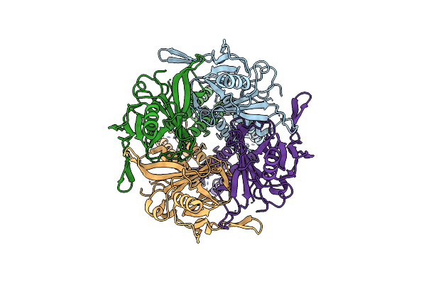
Crystal Structure Of 2-O-Alpha-Glucosylglycerol Phosphorylase In Complex With Glucose
Organism: Bacillus selenitireducens
Method: X-RAY DIFFRACTION Resolution:1.90 Å Release Date: 2014-05-21 Classification: TRANSFERASE Ligands: BGC, 1PE, PG4, CA |
 |
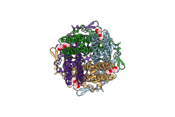
Crystal Structure Of 2-O-Alpha-Glucosylglycerol Phosphorylase In Complex With Isofagomine And Glycerol
Organism: Bacillus selenitireducens
Method: X-RAY DIFFRACTION Resolution:2.30 Å Release Date: 2014-05-21 Classification: TRANSFERASE Ligands: IFM, 1PE, GOL, P6G, PGE, CA, PG4, 7PE, PE4, PE5 |
 |
Crystal Structure Of Beta-Mannanase From A Symbiotic Protist Of The Termite Reticulitermes Speratus
Organism: Symbiotic protist of reticulitermes speratus
Method: X-RAY DIFFRACTION Resolution:1.30 Å Release Date: 2014-03-05 Classification: HYDROLASE Ligands: NAG, NA, MG |
 |
Crystal Structure Of Beta-Mannanase From A Symbiotic Protist Of The Termite Reticulitermes Speratus Complexed With Gluco-Manno-Oligosaccharide
Organism: Symbiotic protist of reticulitermes speratus
Method: X-RAY DIFFRACTION Resolution:1.40 Å Release Date: 2014-03-05 Classification: HYDROLASE Ligands: BMA, BCT, NA, MG |
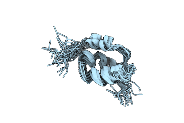 |
Organism: Mus musculus
Method: SOLUTION NMR Release Date: 2013-04-17 Classification: SIGNALING PROTEIN |