
Search Count: 2,138
 |
Organism: Homo sapiens
Method: X-RAY DIFFRACTION Release Date: 2025-12-17 Classification: PROTEIN BINDING |
 |
Organism: Vibrio cholerae o1 biovar el tor str. n16961
Method: X-RAY DIFFRACTION Release Date: 2025-12-10 Classification: HYDROLASE Ligands: NI, BB2 |
 |
Organism: Vibrio cholerae o1 biovar el tor str. n16961
Method: X-RAY DIFFRACTION Release Date: 2025-12-10 Classification: HYDROLASE Ligands: BB2, NI |
 |
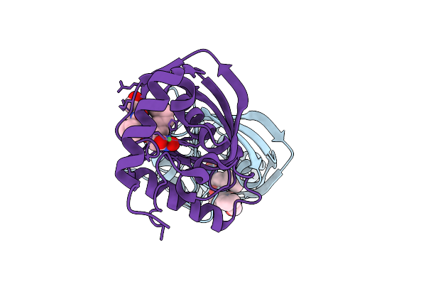
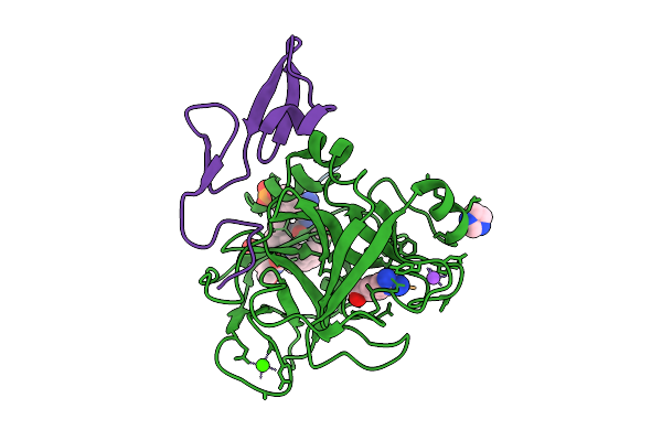
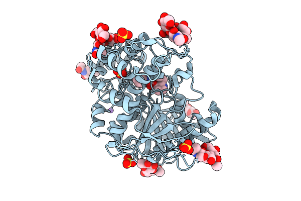
Structure Of Human Neutrophil Elastase In Complex With A 5,8-Dihydro[1,2,4]Triazolo[4,3-A]Pyrimidin-3(2H)-One Inhibitor
Organism: Homo sapiens
Method: X-RAY DIFFRACTION Release Date: 2025-12-10 Classification: HYDROLASE Ligands: NAG, FUC, A1JN2 |
 |
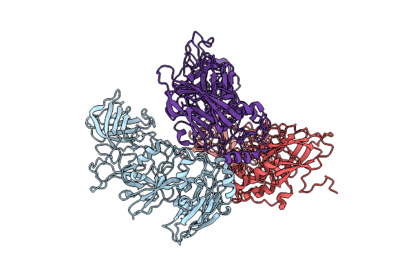
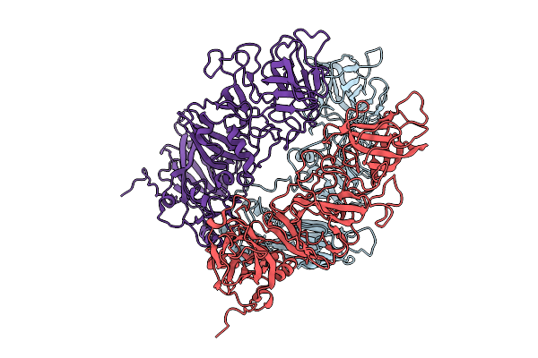
Organism: Rabbit hemorrhagic disease virus
Method: ELECTRON MICROSCOPY Release Date: 2025-12-03 Classification: VIRAL PROTEIN |
 |
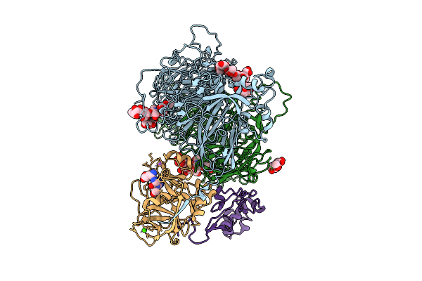
Organism: Homo sapiens, Synthetic construct
Method: X-RAY DIFFRACTION Release Date: 2025-11-26 Classification: BLOOD CLOTTING Ligands: CA, NA, CL, IMD, 0GJ, GLU, ILE, PHE, PTR |
 |
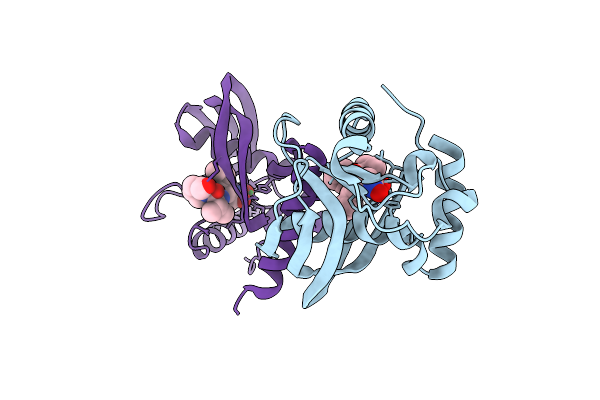
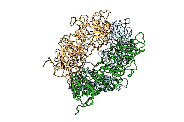
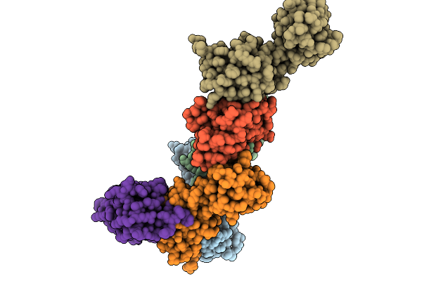
A 3.3 Angstrom Cryo-Em Structure Of An Engineered High-Affinity Human Prothrombinase Complex
Organism: Homo sapiens
Method: ELECTRON MICROSCOPY Release Date: 2025-11-26 Classification: BLOOD CLOTTING Ligands: CA, NA, 0GJ, CU |
 |
Organism: Rabbit hemorrhagic disease virus-ast89, Oryctolagus cuniculus
Method: ELECTRON MICROSCOPY Release Date: 2025-11-26 Classification: VIRUS LIKE PARTICLE |
 |
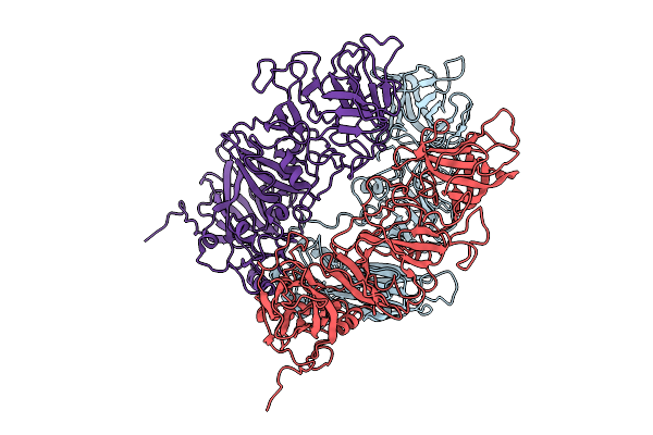
Organism: Rabbit hemorrhagic disease virus
Method: ELECTRON MICROSCOPY Release Date: 2025-11-26 Classification: VIRAL PROTEIN |
 |
Organism: Rabbit hemorrhagic disease virus
Method: ELECTRON MICROSCOPY Release Date: 2025-11-26 Classification: VIRUS |
 |
Organism: Oryctolagus cuniculus
Method: ELECTRON MICROSCOPY Release Date: 2025-11-26 Classification: VIRUS |
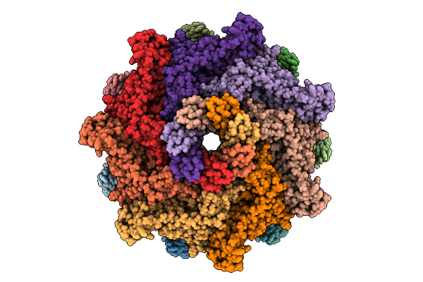 |
Organism: Aeromonas hydrophila
Method: ELECTRON MICROSCOPY Release Date: 2025-11-26 Classification: TOXIN |
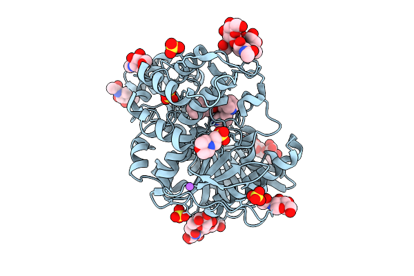 |
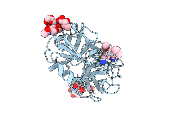
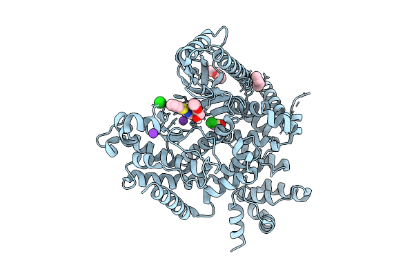
Structure Of Recombinant Human Butyrylcholinesterase In Complex With Naphthalen-2-Yl Methyl((2S,3R)-2-(Pyridin-3-Ylmethyl)Quinuclidin-3-Yl)Carbamate
Organism: Homo sapiens
Method: X-RAY DIFFRACTION Release Date: 2025-11-26 Classification: HYDROLASE Ligands: NAG, A1JOH, MES, SO4, NA |
 |
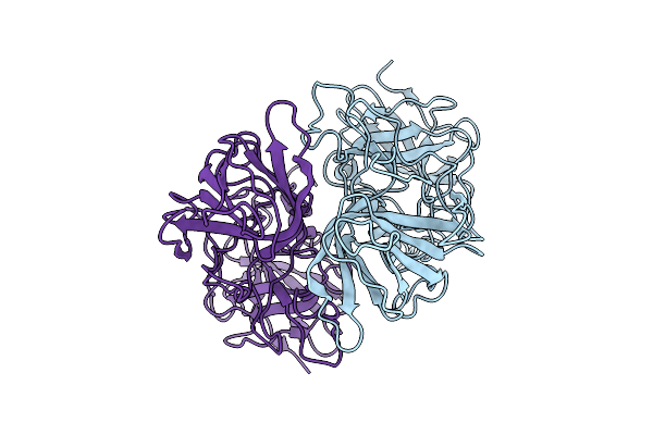
Structure Of Recombinant Human Butyrylcholinesterase In Complex With (2S, 3R)-2-(Pyridin-3-Ylmethyl)Quinuclidin-3-Yl Phenylcarbamate
Organism: Homo sapiens
Method: X-RAY DIFFRACTION Release Date: 2025-11-26 Classification: HYDROLASE Ligands: NAG, A1JOI, SO4, CL, NA |
 |
Organism: Streptomyces lividans 1326
Method: X-RAY DIFFRACTION Release Date: 2025-10-08 Classification: OXIDOREDUCTASE Ligands: HEM |
 |
Organism: Aeromonas hydrophila
Method: ELECTRON MICROSCOPY Release Date: 2025-10-08 Classification: TOXIN |
 |
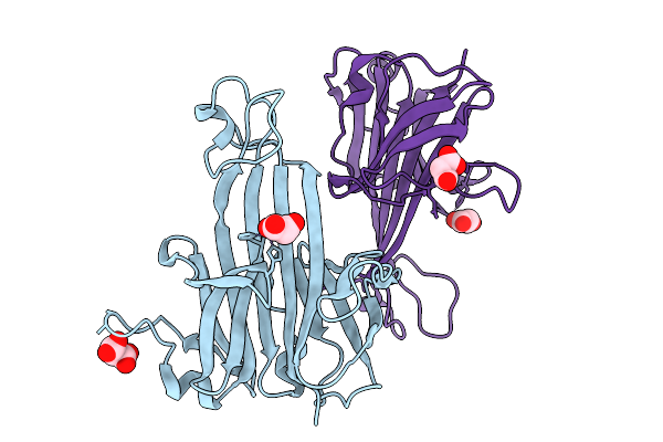
Organism: Homo sapiens, Vicugna pacos
Method: X-RAY DIFFRACTION Release Date: 2025-10-01 Classification: IMMUNE SYSTEM Ligands: EDO, SO4 |
 |
The Structure Of Human Vacuolar Protein Sorting 34 Catalytic Domain Bound To Rd-I-53
Organism: Homo sapiens
Method: X-RAY DIFFRACTION Release Date: 2025-09-17 Classification: TRANSFERASE Ligands: A1A6F, ETX, ETZ, DMS, K, CL |
 |
Organism: Piscinibacter sakaiensis
Method: X-RAY DIFFRACTION Release Date: 2025-09-10 Classification: METAL BINDING PROTEIN Ligands: NI, MLI |
 |
Ang-1 Domain Of The Hupe/Urej-2 Protein From Rhodobacteraceae Bacterium Rbang-1A With Cu Bound
Organism: Rhodobacteraceae bacterium pd-2
Method: X-RAY DIFFRACTION Release Date: 2025-09-10 Classification: METAL BINDING PROTEIN Ligands: ZN, CU |